



Risk Factors Associated with Amelanotic Melanoma Development
Would you recognize a dangerous skin growth if it were pink rather than black? Most people imagine an uneven, dark mole when they think about skin cancer. However, some serious lesions contain little visible pigment and may resemble a pimple, scar, bite, or harmless red patch.
Amelanotic melanoma is uncommon, but its less familiar appearance can delay recognition. The condition develops from pigment-producing cells, yet the tumor creates little or no visible melanin. As a result, color-based warning rules may be less useful.
This article explains the strongest known risk factors, the difference between general and subtype-specific risks, and the changes that deserve a dermatologist’s attention. It also covers sun exposure, age, genetics, immune health, personal history, and practical prevention.
Medical note: Risk factors cannot diagnose a skin lesion. A new, growing, bleeding, or persistent mark should be examined by a qualified healthcare professional.
What Is Amelanotic Melanoma?
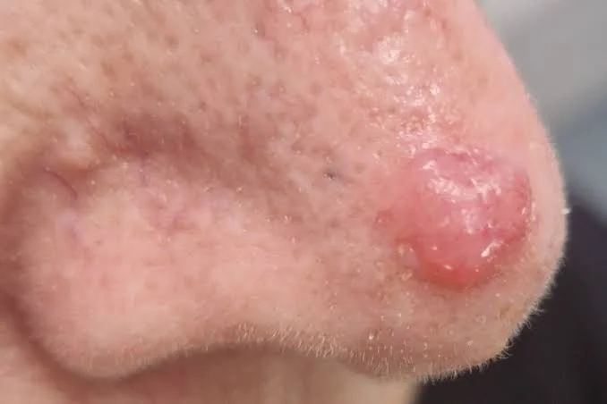
This cancer begins in melanocytes, which are cells that normally produce the pigment melanin. In this form, the abnormal cells make little or no mature pigment, so the lesion may appear red, pink, pale, or skin-colored instead of dark brown or black.
Published estimates suggest that nonpigmented and partly pigmented tumors account for roughly 2% to 20% of cases. The broad range reflects differences in definitions, because some studies include lesions with a small amount of color while others count only completely colorless tumors.
Any major subtype can lack pigment. For example, nodular melanoma may present as a fast-growing pink bump rather than a dark, irregular mole.
The older medical term malignant melanoma may still appear in health records and research papers. However, modern patient information usually uses simpler wording.
Why Risk Factors Matter
A risk factor raises the chance of developing a disease, but it does not guarantee that the disease will occur. Some people have several risk factors and never develop cancer, while others receive a diagnosis without any obvious risk.
It is also important to separate two questions:
- What increases a person’s overall chance of developing this form of skin cancer?
- What makes an existing tumor more likely to have little visible pigment?
Research has clearer answers for general cancer risk than for the loss of pigment itself. Therefore, responsible health advice should avoid claiming that one feature directly causes a nonpigmented tumor.
Major Risk Factors for Amelanotic Melanoma
1. Increasing Age
Older age is one of the most consistent associations. Studies have found that people diagnosed with nonpigmented tumors tend to be older than those with visibly pigmented lesions.
Age does not directly cause cancer. However, DNA damage can build over time, and older adults have usually had more years of ultraviolet exposure. Age-related changes in immune function may also affect the body’s ability to identify and remove abnormal cells.
These lesions are not limited to older people. Younger adults and, rarely, children can also be affected. Consequently, a changing growth should not be dismissed because someone seems “too young.”
2. Long-Term Ultraviolet Exposure
Ultraviolet radiation from sunlight and indoor tanning devices can damage DNA within skin cells. This damage is a well-established risk factor for melanoma skin cancer, as well as other common forms of skin cancer.
Nonpigmented lesions are frequently reported on sun-exposed areas, particularly the face, scalp, neck, arms, and upper body. Chronic sun damage may be especially relevant among older adults.
However, ultraviolet exposure does not explain every case. Tumors can develop on areas that receive little sunlight, including the feet, nails, and covered skin.
3. Severe or Repeated Sunburn
A history of blistering sunburn, especially during childhood or adolescence, increases overall risk. Adult sunburn also matters because every burn represents injury to the skin.
Occasional intense exposure may be particularly important. For example, someone who works indoors but spends holidays tanning without protection may receive a large ultraviolet dose over a short period.
Moreover, a tan is not proof of safety. Tanning is a biological response to cellular injury, even when the skin does not visibly burn.
4. Fair Skin and a Tendency to Burn
People with fair skin, light eyes, red or blond hair, heavy freckling, and skin that burns easily have a higher overall risk. These traits often reflect lower levels of protective melanin and reduced natural defense against ultraviolet radiation.
Research has also connected red hair and severe freckling with a greater likelihood of nonpigmented presentation compared with conventional pigmented tumors.
Still, people with brown or black skin can develop the disease. No skin tone provides complete protection, and unfamiliar-looking changes should be assessed regardless of ethnicity.
5. Red Hair and MC1R Gene Variants
Red hair is commonly associated with variations in a gene called MC1R. This gene helps control the type of pigment the body produces and influences how skin responds to ultraviolet radiation.
Some MC1R variants are linked to increased overall risk, partly independently of visible hair color or skin type. Research has also explored whether pigment-related genes influence why certain tumors lose or fail to produce color.
Having red hair does not mean a person will develop cancer. Instead, it is one piece of a wider risk profile that may justify careful sun protection and regular skin checks.
Personal and Family History
Previous Skin Cancer
A personal history of this disease raises the likelihood of developing another primary tumor. A history of basal cell or squamous cell carcinoma may also signal heavy past ultraviolet exposure and a skin type that is more sensitive to damage.
One study found an association between clinically nonpigmented tumors and a history of nonmelanoma skin cancer. Therefore, people who have already received any skin cancer diagnosis should follow the examination schedule recommended by their dermatologist.
Family History and Inherited Risk
Having a close relative with the disease increases overall risk. The concern becomes greater when several first-degree relatives are affected, when diagnoses occur at younger ages, or when a family has numerous unusual moles.
Rare inherited changes, including variants involving CDKN2A, can create a stronger family tendency. However, genetic testing is not automatically useful for everyone and should be discussed with a dermatologist or genetic counselor.
Interestingly, factors that raise general risk do not always make a tumor more likely to be colorless. Some research found that family history and large numbers of moles were less strongly associated with a nonpigmented presentation than with a pigmented one. This does not make those factors protective; it highlights the complexity of subtype research.
Moles, Freckles, and Skin Patterns
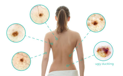
People with many ordinary moles or several atypical moles have an increased overall risk. Atypical moles may be larger, less even, or more varied in color than common moles.
Nevertheless, many tumors arise on previously normal-looking skin rather than from an existing mole. A person should therefore examine the entire skin surface, not only familiar marks.
The “ugly duckling” idea is useful. Most marks on one person tend to share a visual pattern. A spot that looks or behaves differently from the others deserves attention, even when it does not match traditional signs of melanoma.
A Weakened Immune System
The immune system helps identify and control abnormal cells. People who take long-term immunosuppressive medicine after an organ transplant may face a higher risk of several skin cancers.
Immune suppression related to certain diseases or cancer treatments can also increase risk. The exact level depends on the condition, medicine, dose, duration, age, and other personal factors.
People with reduced immunity should ask their medical team how often they need a professional skin examination. They should not stop prescribed medicine without medical guidance.
Previous Radiation and Environmental Exposures
The National Cancer Institute lists certain environmental and occupational exposures among possible risk factors, including previous radiation and contact with some industrial chemicals. However, these associations are less central than ultraviolet exposure, skin type, moles, and personal history.
Risk should be evaluated in context. A brief, low-level exposure does not carry the same meaning as repeated occupational contact over many years.
Where Can a Nonpigmented Lesion Develop?
These tumors can appear almost anywhere. Common sites include the head, neck, upper body, arms, and legs, but hidden areas also matter.
A nonpigmented subungual melanoma may grow beneath a fingernail or toenail without creating the dark streak people expect. The phrase normal black line on nail vs melanoma is commonly searched online, yet a colorless nail-unit tumor may instead appear as nail damage, a red lump, bleeding, or a sore that does not heal.
Likewise, nail melanoma cannot be ruled out simply because no black band is present. Persistent nail lifting, splitting, tissue growth, or unexplained bleeding needs professional assessment.
Ocular melanoma begins inside the eye and has different risk factors, warning features, and diagnostic methods. It should not be confused with a pale growth on the surface of the body.
How Does Amelanotic Melanoma Look?
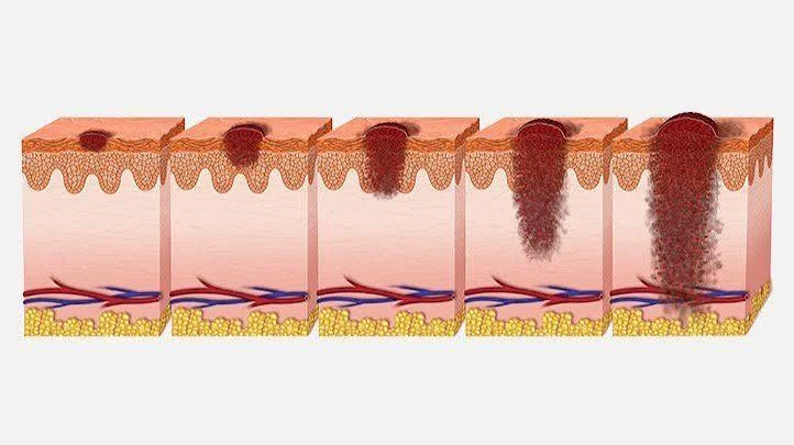
People often search what does melanoma look like on the skin and expect a single reliable picture. In reality, nonpigmented lesions vary greatly.
They may appear as:
- A pink or reddish patch that slowly expands.
- A raised, firm, skin-colored bump.
- A shiny growth resembling basal cell carcinoma.
- A bleeding lump that looks like a pyogenic granuloma.
- A scaly area mistaken for eczema or irritation.
- A sore that repeatedly crusts and reopens.
- A pale lesion with a small brown or grey area near one edge.
The traditional ABCDE rule can miss these changes because color variation may be absent. Dermatologists may also consider the three Rs: red, raised, and recently changing.
Why Diagnosis Can Be Delayed
A pink bump may be treated as acne, a bite, an ingrown hair, or inflammation. A scaly patch may be mistaken for dermatitis, while a bleeding lesion may resemble a harmless blood-vessel growth.
This visual overlap creates a diagnostic challenge for patients and clinicians. Dermoscopy can reveal irregular blood-vessel patterns, faint remaining pigment, white lines, or milky-red areas that are not easy to see with the naked eye.
Still, no visual tool provides complete certainty. A suspicious lesion generally requires removal or biopsy and examination by a pathologist.
Risk Factors Are Not the Same as Prognostic Factors
A risk factor affects the chance of developing a tumor. A prognostic factor helps predict how an existing tumor may behave.
Once a diagnosis is confirmed, important prognostic details include:
- Breslow thickness, or how deeply the tumor has grown.
- The presence or absence of ulceration.
- Involvement of nearby lymph nodes.
- The rate of cell division.
- Spread to distant tissues or organs.
The term metastatic melanoma refers to disease that has moved beyond its original location. However, a pink or colorless appearance does not prove that spread has occurred.
Population research suggests that nonpigmented cases may show poorer outcomes mainly because they are diagnosed at a later stage. When researchers account for stage, survival differences may decrease or disappear.
Can Early Detection Reduce the Risk of Advanced Disease?
Finding early stage melanoma usually allows simpler treatment than finding a thick or widely spread tumor. In many localized cases, surgical removal is the main treatment.
Color should never be the only trigger for concern. A new spot that grows, bleeds, becomes firm, or fails to heal deserves attention even when it matches the surrounding skin.
People sometimes ask, how fast does melanoma spread after a bump appears. There is no universal timetable because growth depends on the tumor’s biology, thickness, subtype, location, and other factors. Rapid visible change should lead to prompt evaluation rather than an attempt to calculate the stage at home.
Practical Ways to Lower Modifiable Risk
Not every risk factor can be changed. Age, natural skin tone, inherited genes, and family history are outside personal control. However, ultraviolet exposure can be reduced.
Useful protective steps include:
- Avoid indoor tanning. Tanning beds expose skin to cancer-causing ultraviolet radiation.
- Use broad-spectrum sunscreen. Choose adequate SPF and reapply it after swimming, sweating, or extended outdoor time.
- Wear protective clothing. Long sleeves, tightly woven fabric, sunglasses, and a broad-brimmed hat add physical protection.
- Seek shade. This is especially useful when sunlight is strongest.
- Protect children carefully. Preventing burns early in life can reduce accumulated damage.
- Check the skin regularly. Include the scalp, ears, back, soles, nails, and areas covered by clothing.
Sun protection lowers avoidable risk, but it cannot guarantee prevention. Anyone can develop a suspicious lesion, including people who have followed careful sun-safety habits.
Personal Insight: Behavior Can Matter More Than Color
A common mistake is waiting for a spot to become dark before taking it seriously. In practice, change is often the more useful clue.
For example, a stable pink mole may remain harmless for many years. By contrast, a new pink bump that doubles in size, bleeds after light contact, and fails to heal deserves examination even though it contains no obvious dark pigment.
The safest approach is not to diagnose the mark from an online gallery. Instead, record when it appeared, photograph it clearly, and describe its changes to a dermatologist.
When Should You Seek Medical Advice?
Arrange an appointment when a lesion:
- It is new and continues to grow.
- Looks different from nearby marks.
- Bleeds without a clear injury.
- Repeatedly crusts or reopens.
- Becomes firm, raised, painful, or itchy.
- Does not improve after suitable treatment.
- Changes noticeably over several weeks.
- Appears beneath a nail and damages the nail plate.
Seek faster care for rapid growth, ongoing bleeding, ulceration, or a growing lump. These changes can have several causes, but guessing can delay the correct treatment.
How Doctors Confirm the Diagnosis
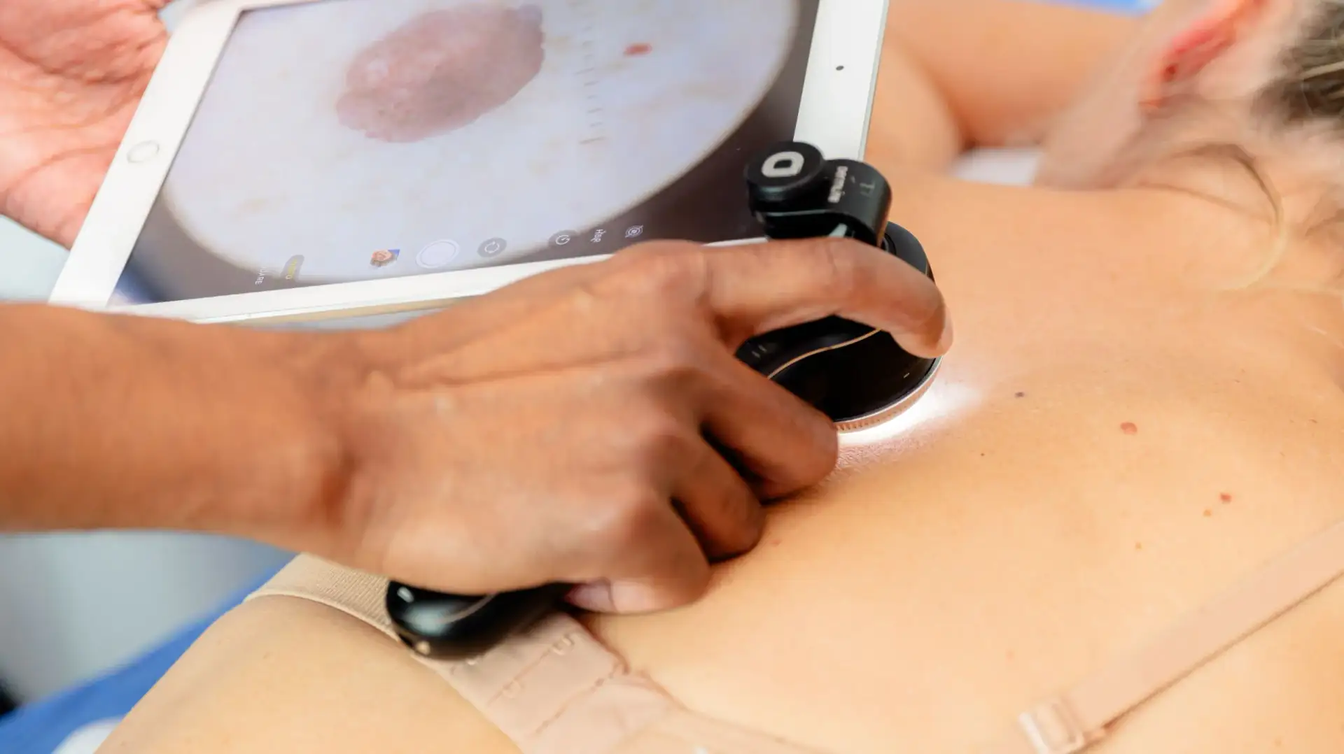
A dermatologist usually examines the lesion and surrounding skin, reviews its history, and may use a dermatoscope. This device magnifies structures and blood vessels below the surface.
When the lesion remains suspicious, a biopsy is required. A pathologist studies the tissue and may use special stains to confirm that the abnormal cells came from melanocytes.
The diagnosis of skin melanoma should never be made from a smartphone photograph alone. Likewise, an app cannot reliably distinguish a harmless pink bump from an unusual cancer in every patient.
Conclusion
The main factors associated with a colorless or lightly colored tumor include increasing age, chronic sun damage, fair skin, severe freckling, red hair, sunburn tendency, certain pigment-related genes, previous skin cancer, and reduced immune function.
However, risk is not destiny. People with no clear risk factors can still be affected, while many high-risk individuals never develop the disease.
The most valuable takeaway is simple: do not rely on dark color as the only warning sign. Watch for spots that are red, raised, recent, growing, bleeding, or different from everything nearby.
A professional examination can provide reassurance when a lesion is harmless and faster treatment when it is not. Paying attention to change is a practical act of prevention, not a reason for panic.
Frequently Asked Questions
Is Amelanotic Melanoma Always Pink?
No. It may be pink, red, pale, flesh-colored, or partly pigmented. Its appearance varies according to location, blood vessels, surface changes, and the small amount of pigment present.
Is It More Dangerous Than Pigmented Disease?
It is often found at a more advanced stage because it is harder to recognize. Research suggests that stage at diagnosis, rather than lack of color by itself, explains much of the difference in outcomes.
Can People with Darker Skin Develop It?
Yes. Every skin tone can be affected. People should seek care for a new, changing, bleeding, or persistent growth regardless of their natural complexion.
Does Every Pink Bump Need a Biopsy?
No. Most pink bumps are not cancerous. However, a dermatologist may recommend biopsy when a lesion grows, bleeds, looks unusual, or cannot be confidently identified during examination.
Can It Develop Without Sun Exposure?
Yes. Ultraviolet exposure is a major modifiable risk, but tumors can arise on covered or low-exposure areas. Genetics, immune health, age, and other biological factors may also play a role.
⭐ Make MindLiva Your Preferred Source on Google
Get trusted health, wellness, and lifestyle articles from MindLiva.
Add MindLiva
Samie Babaei is a medical science student at Shahid Beheshti University of Medical Sciences with a strong interest in health, wellness, and medical research. As a health writer and researcher at MindLiva, she focuses on creating clear, evidence-based content that helps readers better understand health and well-being.