



Early Signs of Nodular Melanoma You Should Never Ignore
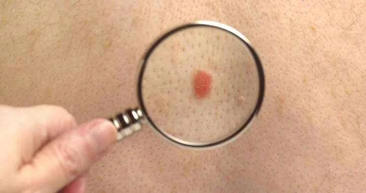
Have you noticed a new bump and assumed it was a pimple, bite, or irritated mole? Most small skin changes are harmless. However, a firm lesion that keeps growing should not be ignored, especially when it looks different from the other marks on your body.
Nodular melanoma matters because it often grows downward into deeper skin sooner than more common flat forms. It may also look smooth, evenly colored, and fairly round, so the familiar ABCDE checklist does not always tell the whole story.
Here, you will learn the main warning features, how doctors investigate suspicious growth, and which changes call for a quick appointment rather than a “wait and see” approach.
Medical note: This article offers general education and cannot diagnose a lesion. A new, changing, bleeding, or persistent growth should be examined by a qualified healthcare professional.
What Is Nodular Melanoma?
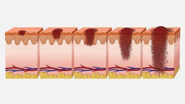
This cancer begins in melanocytes, the cells that make the pigment melanin. Unlike a lesion that spends a long time spreading across the surface, the nodular form commonly develops as a raised growth and can move vertically into the skin relatively early.
It represents about 15% to 20% of cases in some clinical summaries. Although less common than superficial spreading disease, its quicker downward growth may produce a thicker tumor by diagnosis.
Tumor thickness is one factor doctors use to determine the stage and select an appropriate treatment.
The growth may be black, brown, blue-black, red, pink, or close to the person’s skin tone. Some lesions are smooth and shiny, while others become rough, crusted, sore, or ulcerated.
Therefore, color alone cannot confirm or exclude cancer.
Broader terms such as melanoma skin cancer and malignant melanoma describe cancer arising from pigment-producing cells. Only examination and tissue analysis can establish the exact diagnosis.
Why Early Signs of Nodular Melanoma Are Missed

Many people expect a dangerous mole to be flat, irregular, multicolored, and larger than a pencil eraser. Those features are useful, but a nodular growth may be symmetrical, have a clear edge, show one color, and start smaller than 6 millimeters.
This explains why early stage melanoma can escape attention when someone checks only for classic irregular moles. The better question is not simply, “Does it look strange?”
It is also important to ask, “Is it new, firm, raised, or steadily changing?”
Dermatology guidance also uses the “ugly duckling” idea. A growth deserves attention when it looks or behaves differently from the person’s other marks, even when it does not match a textbook image.
The EFG Rule: Elevated, Firm, and Growing
The EFG rule is especially useful for assessing a raised lesion:
- Elevated: The spot rises above nearby skin or becomes thicker.
- Firm: It feels solid rather than soft.
- Growing: It continues to enlarge or change over days, weeks, or a month.
These features do not prove cancer. Cysts, scars, inflamed bites, and other harmless conditions can also feel raised or firm.
However, continuous growth combined with elevation and firmness is a strong reason to arrange a professional assessment.
Seven Warning Changes You Should Check
1. A New Bump That Keeps Enlarging
A new bump is not automatically dangerous, but ongoing growth matters. Photograph it beside a ruler, note the date, and seek advice if it becomes wider or taller.
Do not dismiss a changing lesion simply because it remains small. Some serious growths measure less than 6 millimeters when first noticed.
2. A Firm, Dome-Shaped Growth
This subtype often presents as a firm, rounded, or dome-shaped lump with a smooth, rough, or crusted surface.
A spot that remains after an expected pimple, shaving injury, or insect bite deserves closer attention. Moreover, squeezing or repeatedly scratching it may create irritation and hide meaningful changes.
3. A Dark, Red, Pink, or Skin-Colored Lesion
Some cancers contain obvious dark pigment, but amelanotic melanoma has little or no visible dark coloring. It may appear pink, red, pale, or similar to the surrounding skin.
Therefore, do not use black or brown coloring as the only danger signal. Growth, firmness, bleeding, and persistent change may be more informative than shade alone.
4. Bleeding, Oozing, or Crusting
Repeated bleeding, unexplained oozing, crusting, or an open area that does not settle deserves medical review.
These changes can occur in several conditions, so the goal is professional evaluation rather than guessing the cause at home.
5. Itching, Tenderness, or Pain
Itching, tenderness, burning, or pain can occur, especially in a new or evolving spot.
In short, comfort does not prove safety, and discomfort does not automatically mean cancer. The symptom should be considered together with visible changes and the lesion’s history.
6. A Spot Unlike Everything Else
Most ordinary moles on one person share a general pattern. A spot that stands out in color, height, texture, shape, or growth rate deserves attention.
This principle works across different skin tones because it focuses on personal change rather than one universal image.
7. A Growth in a Hidden Area
Check the scalp, ears, back, buttocks, soles, between the toes, and around the nails.
Cancer can develop in places that receive little direct sunlight, so use mirrors or ask a trusted partner to inspect areas you cannot see clearly.
What Does Nodular Melanoma Look Like?
People often search what does melanoma look like on the skin, expecting one clear picture. In reality, its appearance varies.
One lesion may be a shiny black bump, another may be reddish and crusted, and another may appear almost colorless.
The ABCDE guide still helps:
- A — Asymmetry: One half differs from the other.
- B — Border: The edge is irregular, blurred, or notched.
- C — Color: The spot contains uneven shades.
- D — Diameter: It may exceed 6 millimeters, although smaller lesions can be serious.
- E — Evolving: It changes in size, shape, color, height, feeling, or surface.
However, a nodular lesion may not show several of these traits.
Use ABCDE together with EFG, personal comparison, and the history of change. The most dependable diagnosis comes from removing suspicious tissue and examining it under a microscope, not from matching phone photos with online galleries.
Related Types and Less Obvious Locations
The word melanoma covers cancers beginning in pigment-producing cells, including forms in less obvious locations.
Subungual melanoma develops beneath or around a fingernail or toenail. It may produce a dark band, nail damage, splitting, or pigment that spreads onto nearby skin.
Meanwhile, nail melanoma should be considered when a new band changes, widens, becomes irregular, affects one nail, or extends beyond the nail plate.
The question of the normal black line on nail vs melanoma cannot be answered safely by color alone. Injury, medication, infection, and natural pigmentation can cause nail bands, while suspicious lines may also be subtle.
A dermatologist can examine the pattern with magnification and decide whether monitoring or tissue testing is needed.
Ocular melanoma begins in the eye. The phrase skin melanoma refers to a cutaneous tumor, while metastatic melanoma means cancer has spread from its original site to lymph nodes, organs, or other tissues.
These terms describe different medical situations and should not be treated as interchangeable.
How Fast Can It Grow or Spread?
A common question is how fast does melanoma spread after a suspicious mark appears. There is no single timetable.
Growth varies according to the subtype, tumor biology, location, immune response, and how early the lesion is found.
What makes the nodular form concerning is its tendency to grow downward relatively quickly. Visible change may occur over days or weeks, yet a person cannot determine depth or spread from the surface appearance.
Visible enlargement does not prove that cancer has spread. Likewise, a small surface diameter does not guarantee a shallow tumor.
Doctors use thickness, ulceration, examination findings, and sometimes lymph-node assessment or imaging to evaluate the disease. Therefore, the practical response to rapid growth is prompt evaluation rather than calculating a personal timeline online.
When Should You See a Dermatologist?
Arrange an appointment promptly when you notice:
- A new firm bump that continues to grow.
- A spot changing in size, color, shape, height, or texture.
- Unexplained bleeding, crusting, oozing, itching, or pain.
- A lesion that looks unlike your other marks.
- A sore that repeatedly heals and returns.
- A changing band beneath a nail.
- A growth that remains after an expected injury, bite, or pimple has healed.
These are signs of melanoma awareness, not a self-diagnosis checklist. Many suspicious-looking growths are benign, but a qualified clinician should make that distinction.
Seek faster care when a lesion is changing rapidly, bleeding without a clear injury, or becoming ulcerated. When booking, describe it as new and changing so the clinic can triage the request correctly.
Avoid removing, freezing, burning, or applying harsh chemicals to an unexplained growth. Altering the surface may delay diagnosis and make the lesion more difficult to assess.
How Doctors Confirm the Diagnosis
A clinician reviews the lesion’s history and may examine it with a dermatoscope. This handheld device provides a magnified view of colors and structures that may not be visible to the naked eye.
When cancer is suspected, the key step is usually a biopsy or complete removal for laboratory study. A pathologist examines the cells and measures important features such as tumor thickness and ulceration.
Further testing and treatment depend on the final stage. Some people need surgery around the original site, while others may require lymph-node evaluation, imaging, immunotherapy, targeted treatment, radiation, or another specialist plan.
Photographs and artificial-intelligence apps may help document change, but they cannot replace tissue diagnosis.
A Simple Monthly Skin-Check Routine
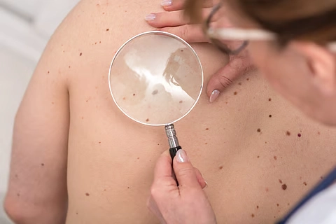
Choose a regular date and examine your skin in a brightly lit room. Remove nail polish so the nail plates and surrounding skin are visible.
Check the following areas:
- Face, ears, scalp, neck, chest, and abdomen.
- Arms, elbows, hands, fingers, and nails.
- Back, shoulders, buttocks, and backs of the legs.
- Feet, soles, heels, toes, and spaces between the toes.
- Areas covered by underwear or normally hidden from sunlight.
Photograph unusual spots consistently, without beauty filters or dramatic lighting. Keep the camera at a similar distance and include a ruler when possible.
A monthly routine is a useful memory aid, although people with a higher risk may need a different schedule from their dermatologist.
Personal Insight: Change Matters More Than a Perfect Match
People often search for an exact visual match. They compare one photograph with dramatic online images and feel reassured when none looks identical.
That approach can create false confidence.
A better habit is to record what is new, what is changing, and what behaves differently from the rest of your skin. A plain-looking bump with steady growth may deserve more attention than a large mole that has remained unchanged for years.
For example, a person may ignore a smooth pink bump because it lacks dark pigment. Yet its steady growth over several weeks may be more important than its color.
Reducing Risk Without Losing Perspective
Reduce avoidable ultraviolet exposure with broad-spectrum sunscreen, protective clothing, shade, and no tanning beds.
Also, learn your normal skin pattern, especially if you have atypical moles, previous skin cancer, heavy ultraviolet exposure, or a strong family history.
Prevention does not replace detection. Even people who protect their skin carefully can develop suspicious lesions, and no one should feel blamed for a diagnosis.
Conclusion
Nodular melanoma may begin as a small, raised, firm bump rather than a classic irregular mole. It can be dark, red, pink, or skin-colored, and it may grow, bleed, itch, crust, or simply look different from everything nearby.
The main takeaway is clear: do not wait for every textbook warning feature to appear.
Use the ABCDE and EFG rules together, photograph meaningful changes, examine hidden areas, and seek professional care for a persistent or growing lesion.
Early action does not mean assuming the worst. It means giving a dermatologist the chance to identify a harmless condition—or address a serious one—without avoidable delay.
Frequently Asked Questions
Can a nodular lesion be small?
Yes. A serious growth can measure less than 6 millimeters. Size is only one clue; continued growth, firmness, elevation, bleeding, and other changes also matter.
Is every firm bump cancerous?
No. Cysts, scars, bites, and benign growths may feel firm. However, a new bump that keeps growing, changes, or bleeds should be assessed rather than diagnosed at home.
Can it be pink or skin-colored?
Yes. Some lesions contain little visible pigment and may look pink, red, pale, or skin-colored. That is why behavior and continued change are as important as color.
Does an itchy mole always mean cancer?
No. Irritation and common skin conditions can cause itching. Still, new itching in a changing spot, particularly with bleeding or growth, deserves medical review.
Can a dermatologist diagnose it by looking?
A visual and dermoscopic examination can show whether a lesion is suspicious. However, laboratory examination of removed tissue is needed to establish the diagnosis.
⭐ Make MindLiva Your Preferred Source on Google
Get trusted health, wellness, and lifestyle articles from MindLiva.
Add MindLiva
Samie Babaei is a medical science student at Shahid Beheshti University of Medical Sciences with a strong interest in health, wellness, and medical research. As a health writer and researcher at MindLiva, she focuses on creating clear, evidence-based content that helps readers better understand health and well-being.