



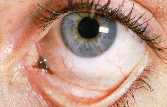
Understanding Ocular Melanoma: Symptoms and Early Signs
Have you ever noticed new flashes, drifting spots, or a change in one eye and wondered whether it matters? Most vision changes have causes far more common than cancer. However, a new or lasting change should not be ignored.
Ocular melanoma begins in pigment-producing cells inside or around the eye. It is rare, and many tumors cause no clear symptoms at first. Therefore, routine dilated eye examinations can matter, especially for people with known pigmented eye lesions or other risk factors.
This guide explains where the disease starts, which symptoms need attention, how specialists confirm a diagnosis, what treatment may involve, and why long-term follow-up is important. The aim is not to create fear. Instead, it is to help readers respond to meaningful changes with calm, informed action.
What Is Ocular Melanoma?
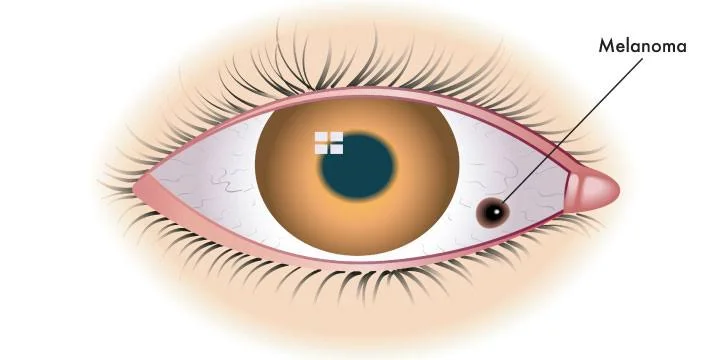
Ocular melanoma develops from melanocytes, the cells that make pigment. Most tumors begin in the uvea, the middle layer of the eye. The uvea includes the iris, ciliary body, and choroid. Most intraocular cases start in the choroid, a blood-rich layer beneath the retina.
It is the most common primary eye cancer in adults, although it remains rare. The American Cancer Society estimates that about 3,200 new cancers of the eye and orbit will be diagnosed in the United States in 2026, with most being eye melanomas.
This disease is biologically different from melanoma skin cancer. Both begin in pigment-making cells, but they often have different genetic changes, patterns of spread, and responses to medicine.
As a result, treatment advice for a skin tumor cannot simply be applied to a tumor beginning inside the eye.
Where Does It Begin?
The main starting sites are:
- Choroid: The most common location, deep in the back of the eye.
- Ciliary body: A hidden area that helps the eye focus and make fluid.
- Iris: The colored part of the eye, where a growing spot may be easier to notice.
- Conjunctiva: The thin tissue covering the white of the eye; tumors here are uncommon and managed differently.
Location affects symptoms, treatment choices, vision, and the chance of spread. For example, a small iris lesion may be noticed sooner than a choroidal growth hidden behind the pupil.
Ocular Melanoma Symptoms to Watch
Many people have no symptoms when the tumor is small. The National Cancer Institute notes that it may be discovered during a routine dilated examination before the patient notices a problem.
Possible symptoms include:
- Blurred or distorted vision
- A missing area in the field of sight
- New floaters, spots, or squiggly lines
- Flashes of light
- A growing dark area on the iris or eye surface
- A change in pupil size or shape
- A change in eye position or movement
- Bulging of one eye
- Sudden or gradual vision loss
These symptoms may also result from retinal problems, migraine, cataracts, inflammation, or normal aging. Still, persistent or sudden changes need examination because appearance alone cannot identify the cause.
Does It Cause Pain?
Pain is not a common early symptom. The American Cancer Society states that pain is rare unless a tumor has grown extensively outside the eye. Therefore, the absence of pain does not rule out a serious problem.
Seek urgent eye care for sudden flashes, a shower of new floaters, a curtain-like shadow, or rapid vision loss. These symptoms may signal a retinal emergency even when cancer is not the cause.
Why Early Ocular Melanoma Can Be Missed
A small tumor in the back of the eye may not change how the eye looks in a mirror. It may also sit outside central vision, allowing reading vision to remain normal for some time.
A dilated examination gives the clinician a wider view of the retina, optic nerve, choroid, and nearby structures. Photographs and scans can document a suspicious area and show whether it changes over time.
People often search for signs of melanoma, but warning patterns depend on where a tumor starts. A changing skin mole and a hidden choroidal mass require different examinations.
Likewise, early stage melanoma is not one single clinical situation. A small localized eye tumor needs an eye-cancer specialist, while an early skin lesion is normally assessed by a dermatologist and confirmed through skin pathology.
Risk Factors and Eye Freckles
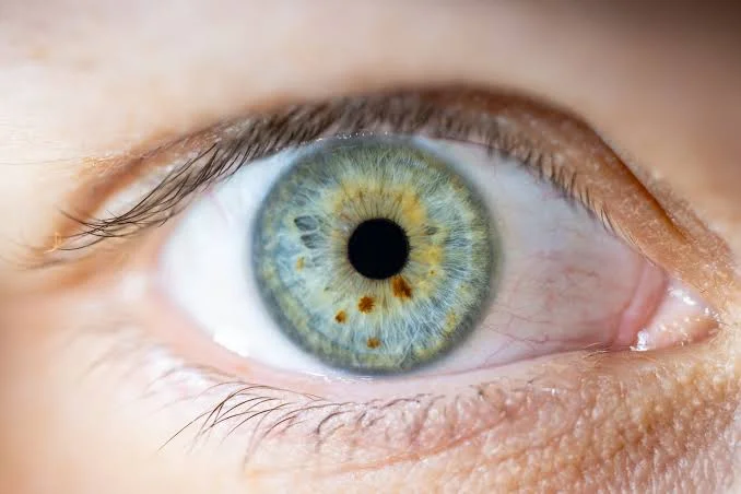
Possible risk factors include older age, fair skin, light-colored eyes, and being White. Certain pigment conditions may also raise risk, although many people who develop the disease have no clear risk factor.
Risk factors do not prove that cancer is present. Moreover, darker eyes or skin do not provide complete protection. Any unexplained eye change deserves appropriate assessment.
A choroidal nevus is a pigmented spot inside the eye, sometimes called an eye freckle. Most remain harmless. However, specialists may monitor thickness, fluid, orange pigment, ultrasound features, and documented growth.
The NCI notes that small uveal tumors can be difficult to distinguish from a nevus. Features such as thickness above 2 millimeters, subretinal fluid, orange pigment, and low internal reflectivity on ultrasound may increase concern.
Monitoring is not neglect. It is an active plan based on repeat imaging at intervals selected for that lesion.
How Ocular Melanoma Is Diagnosed
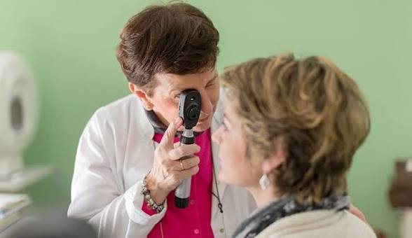
Diagnosis usually begins with a detailed examination by an ophthalmologist, followed when needed by referral to an ocular oncologist. Careful examination by an experienced clinician remains the most important diagnostic step.
Tests may include:
- Dilated ophthalmoscopy to inspect internal eye structures.
- Fundus photography to compare the lesion over time.
- Eye ultrasound to measure thickness and internal features.
- Optical coherence tomography to show retinal detail and fluid.
- Fluorescein angiography to examine blood-flow patterns.
- Body imaging after diagnosis to look for possible spread.
A biopsy is not always required because specialists can often diagnose the tumor through examination and imaging. When tissue is collected, chromosome or gene-expression tests may help estimate the risk of future spread and guide follow-up.
Why Online Photos Are Not Enough
Searches such as what does melanoma look like on the skin or normal black line on nail vs melanoma mainly concern visible body changes, not tumors inside the eye. Likewise, nail melanoma and subungual melanoma require examination of the nail unit.
Other terms may also confuse readers. nodular melanoma usually describes a fast-growing skin form, while amelanotic melanoma refers to a tumor with little visible pigment. skin melanoma is not simply another name for a uveal tumor.
In short, photos may encourage a medical visit, but they cannot replace specialist examination, imaging, or pathology when needed.
Ocular Melanoma Staging and Spread
Doctors consider tumor size, thickness, location, local extension, and whether cancer has reached distant organs. Choroidal and ciliary body tumors may be classified from stage I through stage IV, while iris tumors use a different system.
Because the uvea has a rich blood supply, distant cells usually travel through the bloodstream rather than lymph channels. Systemic spread is found in only about 2% to 3% of patients at diagnosis, but it can appear later. The liver is the most common first site.
People often ask how fast does melanoma spread, but there is no single timeline. Growth depends on genetics, size, location, cell features, and the individual patient.
When an eye tumor reaches distant organs, it is not identical to metastatic melanoma that started in the skin. This distinction matters because drug response and treatment planning can differ.
Treatment Options for Ocular Melanoma
Treatment aims to control the tumor, reduce local complications, preserve the eye when reasonable, and save useful vision whenever possible. The plan depends on size, position, vision potential, general health, and evidence of spread.
Observation
A small, uncertain lesion that is not growing may be watched with repeat photographs, scans, and ultrasound. Observation may also be selected when treatment risks are greater than the likely benefit.
However, follow-up visits must be kept. Documented growth can change the treatment plan.
Radiation Therapy
Radiation is the most common treatment for eye tumors that have not spread. Plaque brachytherapy places a small radioactive disc against the outer wall of the eye near the tumor.
Proton therapy directs radiation from outside the body.
These treatments can often preserve the eye, although vision may decline if radiation affects the retina, optic nerve, lens, or blood vessels.
Surgery
Local surgery may remove selected tumors while leaving the eye in place. Enucleation, or removal of the eye, may be recommended for a very large tumor, severe pain, uncontrolled pressure, optic nerve involvement, or an eye unlikely to retain useful vision.
After enucleation, a patient can usually be fitted with an artificial eye designed to match the appearance of the other eye. Rehabilitation may also help with depth perception, driving, work, and emotional adjustment.
Treatment After Distant Spread
For HLA-A*02:01-positive adults with unresectable or advanced uveal disease, tebentafusp is an approved treatment. It brings immune T cells close to cancer cells.
In its main trial, median overall survival was 21.7 months with tebentafusp compared with 16 months for the control treatments.
Other choices may include immunotherapy, liver-directed procedures, surgery in selected cases, radiation, or clinical trials.
Because advanced uveal disease behaves differently from malignant melanoma of the skin, treatment at an experienced center may be helpful.
Prognosis and Follow-Up
Outlook depends on tumor size, location, genetic features, cell appearance, spread, age, and general health. Statistics describe groups, not one person’s future.
For U.S. patients diagnosed from 2015 through 2021, five-year relative survival was 88% for localized disease, 65% for regional disease, and 19% for distant disease. Newer treatments may improve outcomes beyond what older data show.
Follow-up may include eye examinations, photographs, ultrasound, vision checks, blood tests, and body imaging. The schedule should reflect the tumor’s risk profile and treatment history.
What to Do If You Notice a Change
Use this simple plan:
- Record the symptom: Note when it began and whether it is worsening.
- Check one eye at a time: This may reveal a missing visual area.
- Avoid driving with sudden vision loss: Arrange urgent help.
- Bring a medicine list: Some drugs can affect vision.
- Request specialist review: A suspicious lesion may need ocular oncology.
- Keep follow-up visits: Comparison over time may be essential.
My practical takeaway from current guidance is simple: do not spend days trying to decide whether a visual change is serious enough. A timely dilated examination is safer and more useful than repeated online image searches.
Questions to Ask the Specialist
- Where is the tumor located?
- How large and thick is it?
- Is the diagnosis certain?
- What vision could treatment affect?
- Would molecular testing help?
- How will the liver and other organs be monitored?
- Should my case be reviewed at a specialist center?
- Are clinical trials relevant?
Clear questions help patients compare options and take part in decisions without becoming overwhelmed.
Conclusion
Ocular melanoma is rare, and its first changes may be subtle or completely silent. Blurred vision, flashes, new floaters, pupil changes, or a growing dark spot should be examined, especially when symptoms persist or appear suddenly.
The positive point is that specialists have several ways to diagnose and treat eye tumors while considering cancer control and vision. Moreover, newer systemic treatments have expanded choices for some people with advanced disease.
Stay alert without assuming the worst. Seek professional eye care for unexplained changes, attend follow-up appointments, and ask for clear explanations. Early attention gives the care team the best chance to protect both health and sight.
Frequently Asked Questions
Can ocular melanoma be seen in a mirror?
Sometimes an iris or surface tumor creates a visible dark spot. However, most uveal tumors begin deep inside the eye and cannot be seen without a dilated examination.
Are floaters usually a sign of eye cancer?
No. Floaters are common and often harmless. Still, sudden new floaters, flashes, shadows, or vision loss need prompt assessment because serious retinal problems can cause them.
Can an eye tumor spread after local treatment?
Yes. Local treatment can control the original tumor, but distant disease may appear later in some patients. Long-term eye and medical follow-up may therefore be recommended.
Is a biopsy always required?
No. Specialists can often diagnose a uveal tumor through examination, ultrasound, and other imaging. A biopsy may be useful when the diagnosis is uncertain or genetic information is needed.
Is an eye tumor the same as skin cancer that spreads to the eye?
No. A primary uveal tumor begins in the eye. Cancer that starts elsewhere and reaches the eye is a secondary tumor and may require a different plan.
⭐ Make MindLiva Your Preferred Source on Google
Get trusted health, wellness, and lifestyle articles from MindLiva.
Add MindLiva
Samie Babaei is a medical science student at Shahid Beheshti University of Medical Sciences with a strong interest in health, wellness, and medical research. As a health writer and researcher at MindLiva, she focuses on creating clear, evidence-based content that helps readers better understand health and well-being.