



Understanding Melanoma Skin Cancer and Its Symptoms
Have you ever noticed a new spot and wondered whether it was simply a mole, freckle, or harmless mark? Most skin changes are not dangerous. However, recognizing unusual changes matters because melanoma skin cancer can take several forms and may not always match the dark, uneven mole shown in awareness campaigns.
It can begin on normal-looking skin, inside an existing mole, beneath a nail, or in an area that receives little sunlight. Some growths are brown or black, while others are pink, red, pale, or skin-colored.
In this guide, you will learn what causes the disease, how to recognize possible symptoms, which body areas people often miss, how doctors confirm a diagnosis, and why early medical attention can make such a meaningful difference.
Medical disclaimer: This article provides general information and cannot diagnose a skin lesion. Contact a qualified healthcare professional about any new, changing, bleeding, painful, or persistent spot.
What Is Melanoma Skin Cancer?
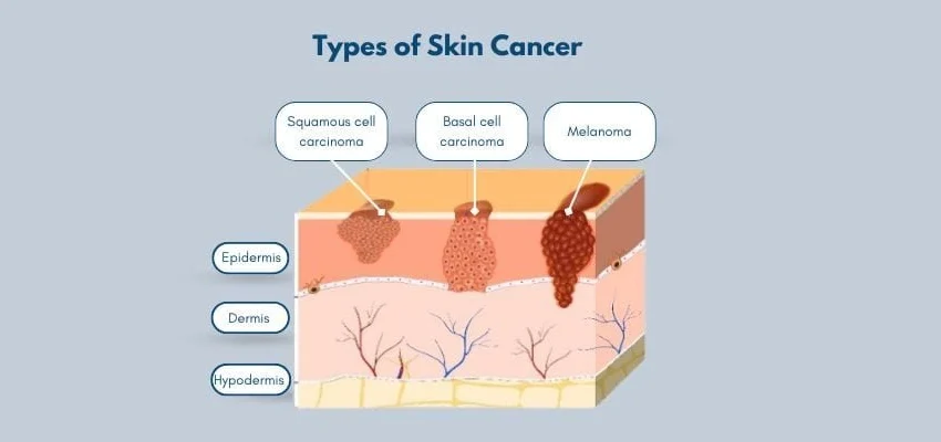
This disease begins when melanocytes, the cells that produce skin pigment, grow without normal control. These cells are found mainly in the skin, although they also exist in the eyes and certain internal tissues.
The condition is less common than basal cell and squamous cell cancers, but it is more likely to invade nearby tissue or spread to other parts of the body when it is not treated early.
The older phrase malignant melanoma may appear in medical records, research papers, and older health articles. Today, healthcare organizations often use simpler language when explaining the condition to patients.
For 2026, the American Academy of Dermatology cites estimates of approximately 112,000 invasive cases and 112,680 in-situ cases in the United States. These figures show why public awareness and early skin checks remain important.
Why Early Detection Matters
When the disease is limited to its original location, treatment is often more straightforward. The American Cancer Society reports a five-year relative survival rate above 99% for localized disease, compared with 76% for regional disease and 35% for distant disease.
These figures describe large groups rather than individual outcomes. Age, general health, tumor biology, treatment response, and other factors can affect a person’s outlook.
Finding early stage melanoma may allow doctors to remove the lesion before it reaches lymph nodes or organs. Therefore, the goal of a skin check is not to diagnose yourself. It is to recognize a meaningful change and arrange professional assessment promptly.
Common Symptoms and Warning Changes
The most important clue is often a new or changing spot. Another useful warning is the “ugly duckling” pattern—a mark that looks different from the others on your body.
Possible signs of melanoma include:
- A mole that changes in size, shape, height, or color
- A new spot that looks unlike nearby marks
- An irregular or poorly defined border
- Several colors within one lesion
- A firm or dome-shaped bump
- Itching, tenderness, pain, or bleeding
- A sore that heals and then returns
- A dark band beneath or around a nail
- A scaly, crusted, or ulcerated surface
Some lesions cause no discomfort. Therefore, the absence of pain does not prove that a growth is harmless.
What Does Melanoma Look Like?
People frequently ask what does melanoma look like on the skin, hoping for one clear description. In reality, its appearance can vary widely.
It may look like a flat brown patch, an uneven mole, a black bump, a red sore, or a pink growth that resembles a cyst. Some lesions are smaller than a pencil eraser when discovered.
Moreover, appearance can vary across different skin tones. In people with darker skin, suspicious growths may be more likely to occur on the palms, soles, or around the nails, although they can develop anywhere.
The ABCDE Warning Guide
The ABCDE method helps people examine moles and other pigmented marks:
- A — Asymmetry: One half does not match the other.
- B — Border: The edge is uneven, blurred, scalloped, or poorly defined.
- C — Color: The lesion contains different shades, such as brown, black, red, white, grey, or blue.
- D — Diameter: Many suspicious lesions are larger than 6 millimeters, but smaller ones can also be serious.
- E — Evolving: The spot changes in color, shape, size, surface, height, or feeling.
The final point—evolution—is especially valuable. A stable mark may be less concerning than a smaller spot that changes noticeably over several weeks.
The Ugly Duckling Sign
Most moles on one person share a general pattern. They may be similar in color, size, or shape.
A mark that looks different from the surrounding group is sometimes called an ugly duckling. Even when it does not meet every ABCDE feature, it should be examined if it is new or changing.
Major Types and Their Different Appearances
Not every tumor grows or looks the same. Doctors classify tumors partly by where they begin and how they develop.
Superficial Spreading Type
This common form often grows outward across the upper skin layers before moving deeper. It may appear flat or slightly raised, with an uneven outline and several colors.
Because its surface may change slowly, photographs can help reveal differences that are hard to remember.
Nodular Type
Nodular melanoma usually appears as a raised, firm growth and may grow downward sooner than some flatter forms. It can be black, brown, blue, red, pink, or close to the surrounding skin color.
Dermatologists sometimes use the EFG guide for a suspicious nodule: elevated, firm, and growing. A bump that continues to enlarge should not be dismissed simply because it is round or evenly colored.
Colorless or Lightly Pigmented Type
Amelanotic melanoma produces little visible pigment. It may resemble a pink pimple, scar, insect bite, irritated patch, or non-healing sore.
This form shows why color should never be the only screening clue. Growth, bleeding, firmness, and continued change may provide more useful information.
Acral and Nail-Unit Types
Acral tumors can develop on the palms, soles, fingers, toes, or nail areas. They are not limited to people with darker skin, although these locations account for a larger share of diagnoses in some darker-skinned populations.
Subungual melanoma begins within the nail unit and can create a brown or black lengthwise streak. It may also cause splitting, lifting, bleeding, nearby skin discoloration, or a lump beneath the nail.
When considering a normal black line on nail vs melanoma, look at whether the band is new, widening, irregular, or spreading onto the cuticle. A bruise usually moves toward the tip as the nail grows, while pigment produced in the nail matrix may remain connected to the base.
The term nail melanoma is sometimes used broadly for suspicious cancer involving the nail apparatus. Any unexplained band or structural change that continues should be assessed by a dermatologist.
Eye and Internal Forms
Ocular melanoma begins in pigment-producing cells inside or around the eye. It differs from the more familiar skin form and requires assessment by an eye specialist.
Rare tumors may also arise on mucosal surfaces, such as the mouth or genital area. These hidden locations demonstrate why not every case is directly linked to sun-exposed skin.
What Causes the Disease?
Cancer develops when genetic changes allow abnormal cells to multiply and survive when they should not. Ultraviolet radiation from sunlight and indoor tanning is a major preventable source of DNA damage in skin cells.
Every exposure does not lead to cancer. The body repairs much of the damage. However, repeated or intense exposure can allow harmful changes to build over time.
Important risk factors include:
- Repeated exposure to natural or artificial ultraviolet light
- A history of severe or blistering sunburns
- Fair skin that burns or freckles easily
- Red or blond hair and light-colored eyes
- Many ordinary moles or several atypical moles
- A personal history of skin cancer
- A close family history of the disease
- Increasing age
- A weakened immune system
- Rare inherited conditions that reduce DNA repair
Anyone can still be affected, including people without obvious risk factors and people of every skin tone. Having several risks does not guarantee a diagnosis, while having none does not provide complete protection.
How Fast Can It Grow or Spread?
The search question how fast does melanoma spread does not have one timetable that applies to everyone. Growth depends on the subtype, tumor thickness, location, genetics, immune response, and other biological factors.
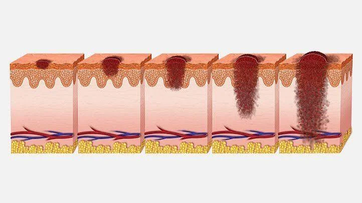
Some lesions change slowly across the skin’s surface. Others develop greater depth more quickly. Visible size alone cannot reveal whether abnormal cells have reached nearby tissue.
Doctors use Breslow thickness to measure how deeply the main tumor has entered the skin. They also consider ulceration, lymph-node involvement, nearby deposits, and distant spread when assigning a stage.
The phrase metastatic melanoma means that cancer cells have moved from the original site to distant tissue or organs. It does not describe every large, dark, or rapidly growing mole.
How Doctors Diagnose a Suspicious Spot
A dermatologist begins by asking when the change appeared, how it has developed, whether it causes symptoms, and whether the patient has relevant personal or family risks.
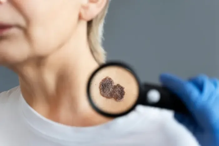
The clinician may use a dermatoscope, a handheld magnifying device that reveals structures beneath the surface. It can help distinguish an ordinary mole from a lesion that requires testing.
However, visual assessment alone cannot provide a final diagnosis. A suspicious area is usually removed or sampled through a biopsy. A pathologist then examines the tissue under a microscope.
The report may include:
- Tumor type
- Breslow thickness
- Ulceration
- Cell-division activity
- Whether the biopsy edges contain abnormal cells
- Other features that help guide staging and treatment
Depending on the findings, doctors may recommend a wider excision, lymph-node evaluation, imaging, or additional laboratory testing.
Understanding the Stages
Staging describes how far the disease has progressed.
- Stage 0: Abnormal cells remain within the outermost skin layer.
- Stage I: The tumor is localized and generally thin.
- Stage II: The tumor remains localized but is thicker or has other higher-risk features.
- Stage III: Cancer has reached nearby lymph nodes, lymphatic channels, or nearby skin.
- Stage IV: Cancer has spread to distant organs or tissues.
This summary is simplified. Formal staging depends on detailed tumor, lymph-node, and metastasis information, so patients should ask their medical team to explain their individual pathology report.
Treatment Options
Treatment depends on stage, tumor biology, location, general health, and previous care.
Surgery
Surgical removal is the primary treatment for many localized tumors. Doctors remove the growth with a margin of normal-looking tissue to reduce the chance that abnormal cells remain.
A sentinel lymph-node biopsy may be discussed when tumor features suggest a meaningful risk of lymphatic spread.
Immunotherapy
Immunotherapy helps the immune system recognize and attack cancer cells. It may be used after surgery in higher-risk cases or to treat advanced disease.
These medicines have improved outcomes for many patients, but they can cause immune-related side effects that require careful monitoring.
Targeted Therapy
Some tumors contain changes in genes such as BRAF. Targeted drugs can block signals that help these cancer cells grow.
Biomarker testing helps doctors decide whether this treatment is suitable. Targeted therapy is not appropriate for every patient.
Radiation and Other Care
Radiation may be used in selected situations, including symptom control or treatment of certain areas. Clinical trials may also offer access to new combinations, vaccines, or treatment approaches.
Current treatment options include surgery, immunotherapy, targeted therapy, radiation, and, less commonly, chemotherapy.
A Practical Monthly Skin Check
Choose a well-lit room and use a full-length mirror together with a hand mirror.
Check:
- The face, lips, ears, scalp, and neck
- The chest, abdomen, sides, and back
- The arms, hands, fingers, and nails
- The buttocks, genital area, and backs of the legs
- The feet, soles, heels, toes, and spaces between them
Take clear photographs of spots you want to track. Use the same lighting and distance, and place a ruler beside the lesion when possible.
Do not rely on memory alone. Small changes become easier to identify when images are compared over time.
Personal Insight: Change Is Often More Useful Than Perfection
Many people wait for a spot to match an alarming internet photograph. This can create false reassurance because real lesions vary in shape, color, and size.
A more useful question is: Has this spot changed from its usual pattern?
For example, a large mole that has remained stable for years may be less urgent than a tiny mark that recently became raised, uneven, itchy, or darker. Likewise, a pink bump that keeps bleeding deserves attention even without dark pigment.
Online images can build awareness, but they cannot diagnose an individual lesion.
Prevention and Risk Reduction
Not every case can be prevented. However, reducing unnecessary ultraviolet exposure can lower avoidable risk.
- Seek shade when sunlight is intense.
- Wear long sleeves, sunglasses, and a wide-brimmed hat.
- Apply broad-spectrum, water-resistant SPF 30 or higher.
- Reapply sunscreen after swimming, sweating, or prolonged exposure.
- Avoid indoor tanning beds and sunlamps.
- Protect children from sunburn.
- Schedule professional examinations based on personal risk.
Sunscreen should support, not replace, shade and protective clothing. Moreover, people with darker skin still need awareness because serious growths can develop in less sun-exposed locations.
When Should You Contact a Dermatologist?
Arrange an appointment for any lesion that:
- Is new and continues to grow
- Changes in size, shape, texture, height, or color
- Looks different from your other marks
- Bleeds without a clear injury
- Itches, hurts, crusts, or repeatedly opens
- Appears as a changing nail streak
- Does not heal after several weeks
Seek faster attention when a spot changes rapidly or develops a firm lump, open surface, or repeated bleeding.
The term skin melanoma should only be applied after proper medical evaluation. Removing, freezing, burning, or treating an unexplained growth at home can delay the correct diagnosis.
Conclusion
Melanoma can appear as a changing mole, firm bump, pink growth, dark nail band, or sore that does not heal. It may begin in a visible area or somewhere easy to overlook.
The ABCDE guide, the ugly duckling sign, and regular full-body checks can help you notice changes sooner. However, no checklist, photograph, or mobile app can replace a professional examination and biopsy when required.
The most important takeaway is simple: pay attention to what is new, different, or changing. Seeking medical advice does not mean assuming the worst. It gives you the best chance to receive reassurance—or timely care—based on expert evaluation.
Frequently Asked Questions
Can Melanoma Be Completely Flat?
Yes. Some lesions remain flat or only slightly raised, especially during earlier surface growth. Any changing patch or mole should be assessed even when it does not form a lump.
Can It Be Pink Instead of Black?
Yes. Some tumors contain little visible pigment and may appear pink, red, pale, or skin-colored. Continued growth, bleeding, or failure to heal can be more important than color.
Does Every Changing Mole Mean Cancer?
No. Moles can change for harmless reasons, including irritation and hormonal shifts. Still, a noticeable or continuing change should be examined rather than diagnosed at home.
Can People with Dark Skin Develop It?
Yes. People of every skin tone can be affected. In darker skin, suspicious areas may occur on the palms, soles, toes, fingers, or around the nails, although any body area can be involved.
Is an Early Diagnosis Curable?
Localized disease is often highly treatable, commonly through surgical removal. The individual outlook depends on tumor depth, ulceration, spread, general health, and response to care.
⭐ Make MindLiva Your Preferred Source on Google
Get trusted health, wellness, and lifestyle articles from MindLiva.
Add MindLiva
Samie Babaei is a medical science student at Shahid Beheshti University of Medical Sciences with a strong interest in health, wellness, and medical research. As a health writer and researcher at MindLiva, she focuses on creating clear, evidence-based content that helps readers better understand health and well-being.