



Vulvar Melanoma vs. Other Skin Cancers: Key Differences
Have you noticed a new dark patch, lump, sore, or changing area on the vulva and wondered whether it needs medical attention? Most vulvar changes are caused by irritation, infection, hormonal changes, or noncancerous skin conditions. However, a lasting or evolving lesion should never be ignored.
Vulvar melanoma is a rare cancer that begins in pigment-producing cells in the vulvar area. It differs from common vulvar squamous cell cancer in its biology, staging, pattern of spread, and treatment response. Moreover, it may be dark, red, pink, or nearly colorless.
This article explains its early symptoms, how it compares with other vulvar and cutaneous cancers, why biopsy matters, and which modern treatment options specialists may consider. It also offers practical steps for checking unfamiliar changes without encouraging self-diagnosis.
What Is Vulvar Melanoma?
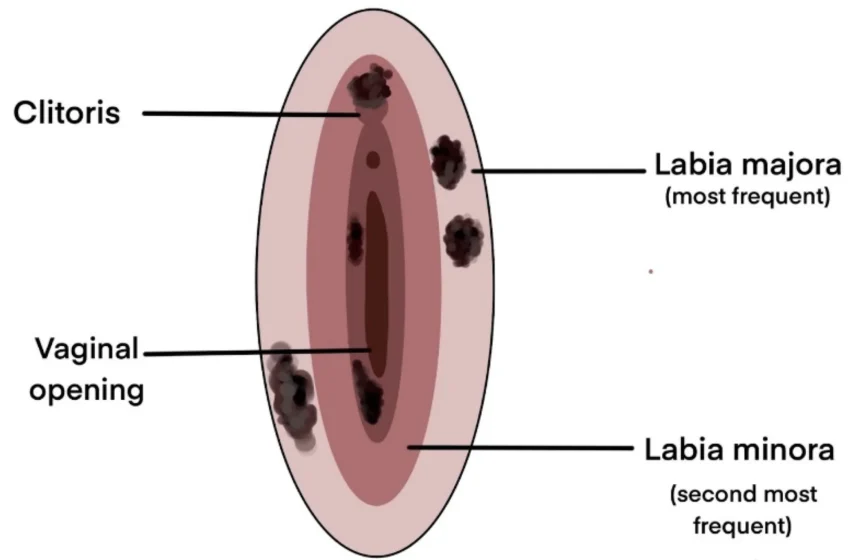
This rare tumor begins in melanocytes, the cells that produce pigment. It may develop on the labia majora, labia minora, clitoral area, or skin near the vaginal opening.
Although the vulva contains both skin and mucosal tissue, these tumors are often grouped with mucosal cancers because many arise on areas that are not normally exposed to sunlight. Their behavior and genetic patterns can differ from those of common sun-related tumors.
A recent expert review published in 2026 described vulvar and vaginal tumors as uncommon diseases for which surgery remains the preferred primary approach. The authors also stressed that evidence is limited because patients have rarely been included in dedicated clinical trials.
How Rare Is It?
Published research estimates that this disease represents roughly 2.4% to 10% of vulvar cancers, with an annual incidence of approximately 0.48 to 1.4 cases per million women. Exact estimates vary because studies cover different countries, periods, and diagnostic classifications.
Another review found that about 10% of women have some type of pigmented vulvar lesion. Fortunately, most of these spots are benign. Therefore, finding a dark mark does not automatically mean cancer, but an unexplained change still deserves examination.
Vulvar Melanoma vs Other Vulvar Skin Cancers
The vulva can develop several types of cancer. They may cause similar symptoms, yet they begin in different cells and require different treatment plans.
Squamous Cell Carcinoma
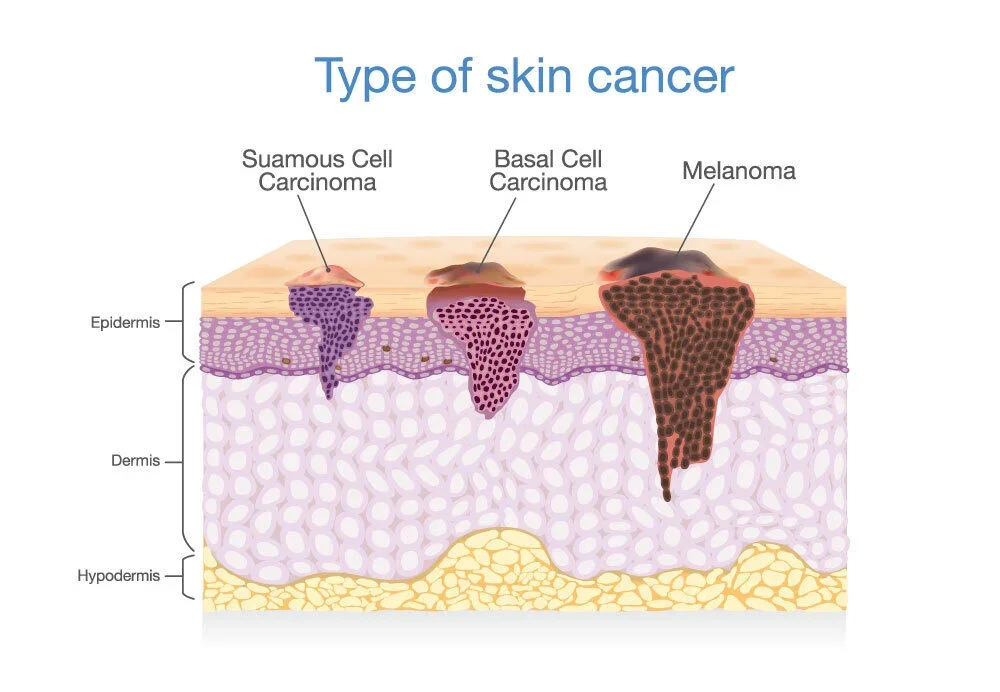
Squamous cell carcinoma is by far the most common vulvar cancer. It begins in the flat cells covering the vulvar surface and makes up the large majority of diagnosed cases.
Some squamous tumors are related to high-risk human papillomavirus infection. Others are associated with chronic inflammatory skin disease and changes involving the TP53 pathway, especially in older patients.
In comparison, vulvar melanoma starts in pigment-making cells and is not usually treated as an HPV-driven disease. Its staging also differs: the standard FIGO system for vulvar carcinoma does not apply, and specialists generally stage it using systems developed for cutaneous tumors.
Basal Cell Carcinoma
Basal cell carcinoma is common on sun-exposed areas of the body but rare on the vulva. It often grows slowly and is less likely to spread to distant organs than a pigment-cell tumor.
It may appear as a sore, firm bump, ulcer, or area of irritation. Because appearance alone is unreliable, a biopsy is still needed to identify the cell type.
Extramammary Paget Disease
Vulvar Paget disease often appears as a red, itchy, scaly, or eczema-like patch. It may be mistaken for dermatitis, a yeast infection, or another chronic skin problem.
Unlike a pigment-cell cancer, Paget disease begins in gland-related cells within the skin. Symptoms may continue for months because creams can briefly reduce irritation without treating the underlying disease.
A Simple Comparison
| Feature | Vulvar Pigment-Cell Tumor | Squamous Cell Cancer | Paget Disease |
| Cell of origin | Melanocyte | Squamous surface cell | Gland-related skin cell |
| Common appearance | Dark, mixed-color, pink, red, or ulcerated lesion | Lump, wart-like growth, sore, or ulcer | Red, scaly, eczema-like patch |
| HPV connection | Not considered a usual driver | Present in many cases | Usually not the main driver |
| Standard staging | Melanoma-based staging | FIGO vulvar cancer staging | Depends on invasion and associated cancer |
| Main diagnostic test | Biopsy | Biopsy | Biopsy |
| Common first treatment | Local surgical removal | Surgery, sometimes radiation or chemoradiation | Surgery or other specialist treatment |
These patterns are useful for understanding differences, but they cannot identify a lesion at home.
Early Signs of Vulvar Melanoma
Some tumors are found during a routine gynecologic or dermatologic examination. Others cause itching, bleeding, pain, a lump, or a visible area of changed color.
General vulvar cancer warning symptoms include a wart-like growth or ulcer, lasting itching, bleeding unrelated to menstruation, and vulvar pain. These symptoms can also be caused by noncancerous conditions.
Possible warning changes include:
- A new brown, black, blue, red, pink, or white patch
- Several colors within one lesion
- A spot with an uneven or blurred edge
- A mole that becomes wider, thicker, or raised
- A sore that does not heal
- Unexplained bleeding or discharge
- Persistent itching, burning, or tenderness
- A firm lump beneath or beside a pigmented area
- A lesion that looks different from nearby spots
- Enlarged groin lymph nodes
A dark color is not required. amelanotic melanoma produces little visible pigment and may resemble a pink bump, raw patch, ulcer, or area of inflamed tissue.
The ABCDE Guide

The familiar ABCDE rule can help identify a suspicious pigmented lesion:
- A — Asymmetry: One half does not resemble the other.
- B — Border: The edge is uneven, notched, or difficult to define.
- C — Color: Several shades appear in the same area.
- D — Diameter: The spot is growing or unusually large.
- E — Evolving: Its appearance or symptoms are changing.
Evolution is especially important. A stable spot may still require assessment, but a lesion that changes in size, color, shape, texture, or feeling deserves prompt attention.
Why Diagnosis Is Often Delayed
The vulva is not an area many people examine regularly. In addition, embarrassment, limited awareness, and the assumption that itching must be an infection can delay an appointment.
Symptoms may also resemble common conditions such as dermatitis, friction, vulvovaginal infection, lichen sclerosus, an ingrown hair, or a harmless mole. A clinician may initially try treatment for one of these conditions when the lesion does not look clearly suspicious.
However, continuing the same treatment without improvement can create further delay. A persistent pigmented lesion, unexplained ulcer, or growth that does not respond as expected should be reassessed.
My strongest practical insight is this: you do not need to decide whether a vulvar spot is dangerous before seeking care. Your role is to notice persistence or change. Diagnosis belongs to a qualified gynecologist, dermatologist, gynecologic oncologist, or pathologist.
How Doctors Diagnose Vulvar Melanoma
Diagnosis begins with a health history and a careful examination of the entire vulva. The clinician may also check the vagina, cervix, nearby skin, and groin lymph nodes.
A magnifying device called a dermatoscope may help reveal pigment structures, vessels, and color patterns that are difficult to see with the naked eye. However, dermoscopy only guides the level of concern.
Biopsy Provides the Answer
A biopsy removes cells or tissue so a pathologist can examine them under a microscope. It is the key test for distinguishing a pigment-cell tumor from a benign mole, squamous cancer, Paget disease, inflammation, or another condition.
The biopsy should be deep enough to measure invasion accurately. Tumor thickness, ulceration, surgical margins, cell division, and other microscopic findings help the team estimate risk and plan treatment.
When the diagnosis is confirmed, additional tests may include:
- A complete skin and mucosal examination
- Examination of groin lymph nodes
- MRI or CT imaging
- PET scanning in selected cases
- Sentinel lymph-node mapping
- Molecular testing of tumor tissue
Imaging is used according to the tumor’s stage, symptoms, and risk features rather than automatically for every small lesion.
How Staging Differs from Other Vulvar Cancers
Most vulvar squamous cancers are staged with the FIGO system. In contrast, the NCI states that FIGO staging does not apply to a malignant melanoma of the vulva; it is staged like its cutaneous counterpart.
Important staging features may include:
- Breslow thickness
- Ulceration
- Nearby tissue involvement
- Regional lymph-node findings
- Satellite or in-transit deposits
- Distant organ spread
The term early stage melanoma generally refers to disease confined to the original area, with no confirmed lymph-node or distant spread. However, even a small vulvar lesion needs careful pathology because depth can be more important than surface width.
Treatment Options
Because the disease is rare, treatment should ideally be planned by a multidisciplinary team that includes gynecologic oncology, dermatology, surgical oncology, medical oncology, pathology, and radiation oncology.
Wide Local Excision
Surgery is the main treatment for a removable primary tumor. Modern care generally aims for complete removal with clear microscopic margins while avoiding unnecessarily radical surgery.
Older treatment sometimes involved radical vulvectomy. However, current reviews support a more tissue-preserving approach when clear margins can be achieved without removing major pelvic organs. The exact margin depends on thickness, location, anatomy, and pathology findings.
This approach can reduce effects on wound healing, urination, movement, sexual function, and body image. Still, a wider operation may be necessary when the tumor involves nearby structures.
Sentinel Lymph-Node Biopsy
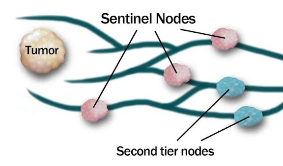
A sentinel lymph-node procedure identifies the first groin node or nodes most likely to receive draining cancer cells. It may help stage selected patients whose lymph nodes do not feel enlarged.
NCI guidance notes that sentinel-node biopsy has shown feasibility and accuracy in vulvar disease, although evidence is more limited than it is for common cutaneous cases. Clinically enlarged or clearly involved nodes require a different evaluation and treatment plan.
Immunotherapy
For high-risk, recurrent, unresectable, or metastatic melanoma, clinicians may consider immune checkpoint inhibitors. These medicines help immune cells recognize and attack tumor cells.
Options used in broader oncology practice include PD-1 inhibitors and combinations that block more than one immune checkpoint. However, response rates in mucosal tumors are generally lower than those reported in many cutaneous trials, and vulvar-specific evidence remains limited.
Targeted Treatment
Molecular testing is important because these tumors may carry changes in genes such as KIT, NRAS, or BRAF. Their mutation pattern is not identical to ordinary sun-related skin melanoma.
A clinically meaningful KIT alteration may support treatment with a KIT-targeting drug in advanced disease. A suitable BRAF change may create other targeted options, although common BRAF V600 mutations are less predictable in mucosal tumors.
Therefore, testing should examine the exact mutation rather than simply report that a gene is “positive.”
Radiation and Clinical Trials
Radiation may be used when surgery cannot remove all disease, when local control is needed, or when cancer has returned. It may also help relieve symptoms from tumors in other body areas.
Because dedicated trials remain scarce, recent expert reviews recommend discussing clinical-trial participation whenever suitable. Trials may study new immunotherapy combinations, targeted drugs, treatment before surgery, or approaches based on tumor biomarkers.
Does Vulvar Melanoma Spread Differently?
Pigment-cell tumors can spread through lymph channels to groin nodes or through the bloodstream to distant organs. Tumor thickness, ulceration, lymph-node involvement, and advanced stage are important risk factors.
Published reviews report poorer average survival than for common cutaneous disease, partly because vulvar tumors may be discovered at a greater depth and may have different biology. One review reported an overall five-year survival estimate of about 47%, compared with 92% for cutaneous disease in the datasets it analyzed. These historical group figures cannot predict an individual outcome.
People often ask how fast does melanoma spread, but no single number applies. Some lesions remain localized for a period, while others behave aggressively. Only pathology, staging tests, and follow-up can estimate a specific person’s risk.
Avoid Confusing Different Melanoma Terms
Online searches often combine cancers that begin in very different places. nodular melanoma usually describes a rapidly growing form on the body’s outer skin, while ocular melanoma begins in or around the eye.
Likewise, subungual melanoma develops in the nail unit, and nail melanoma is a broader public term for tumors affecting that area. A search for normal black line on nail vs melanoma may be useful for nail awareness but does not explain a vulvar lesion.
The phrase melanoma skin cancer commonly refers to cutaneous disease. Searches for what does melanoma look like on the skin or signs of melanoma may show useful examples, but genital lesions often require a different level of privacy, lighting, anatomy knowledge, and specialist examination.
In short, pictures may motivate someone to seek care, but they cannot confirm the diagnosis or identify the exact subtype.
How to Check the Vulvar Area Safely
A self-check is not a substitute for a medical examination. However, it can help you become familiar with your normal skin.
Use a handheld mirror in a private, well-lit room. Look at the outer and inner labia, the clitoral hood, the skin around the vaginal opening, and the nearby perineal area.
Notice:
- New spots or lumps
- Areas with mixed colors
- Sores, cracks, or ulcers
- Thickened or raised tissue
- Unexplained bleeding
- Lasting redness or pale areas
- One-sided swelling
- Changes that remain after an infection treatment
Do not scrub, cut, bleach, freeze, or apply strong acids to a lesion. These actions can injure sensitive tissue and make later examination more difficult.
When to Arrange an Appointment
Book a medical assessment when a vulvar change:
- Is new and unexplained
- Continues for several weeks
- Changes in color, shape, or thickness
- Bleeds without a clear reason
- Becomes painful, itchy, or ulcerated
- Returns after appearing to heal
- Does not improve with prescribed treatment
- Looks clearly different from nearby tissue
Seek faster care for heavy bleeding, severe pain, rapidly increasing swelling, fever, or difficulty urinating.
A useful appointment description might be: “This dark patch appeared three months ago, has become wider, and now bleeds after light contact.” Specific details can help the clinic judge urgency.
Questions to Ask the Specialist
- What conditions could cause this lesion?
- Do I need a biopsy?
- Which type of biopsy will protect accurate staging?
- Should the pathology be reviewed by a specialist?
- What is the tumor’s Breslow thickness?
- Are the surgical margins clear?
- Is sentinel-node testing appropriate?
- Should the tumor undergo KIT, BRAF, and NRAS testing?
- Would a second opinion or clinical trial be useful?
- How will treatment affect sexual function, urination, and healing?
These questions support informed decisions without replacing medical advice.
Conclusion
Vulvar melanoma differs from more common vulvar cancers in its cell of origin, staging, molecular profile, and treatment response. It may appear dark and uneven, but it can also look pink, red, pale, ulcerated, or similar to an irritated patch.
Squamous cancer, Paget disease, benign moles, infections, and inflammatory disorders can produce overlapping symptoms. Therefore, appearance alone is not enough. A properly performed biopsy provides the diagnosis.
The most important takeaway is not to panic over every spot. Instead, respect changes that persist, grow, bleed, itch, hurt, or fail to respond to treatment. Prompt specialist care can provide reassurance when a lesion is harmless and faster treatment when it is not.
Frequently Asked Questions
Is vulvar melanoma always dark?
No. Many lesions contain brown, black, blue, or mixed pigment, but some are pink, red, pale, or almost colorless. Any persistent evolving lesion deserves assessment.
Is it related to HPV?
It is not generally considered an HPV-driven cancer. HPV is much more closely associated with many vulvar squamous cell carcinomas.
How is it diagnosed?
A clinician examines the lesion and performs a biopsy. A pathologist then identifies the cell type and measures features such as depth and ulceration.
Can it be cured with surgery?
Localized disease may be treated successfully with complete surgical removal. The outlook depends on depth, ulceration, margins, lymph nodes, and whether spread is present.
Should every vulvar mole be removed?
No. Many pigmented vulvar spots are benign. However, a new, changing, irregular, bleeding, or symptomatic lesion should be examined before deciding whether removal is necessary.
⭐ Make MindLiva Your Preferred Source on Google
Get trusted health, wellness, and lifestyle articles from MindLiva.
Add MindLiva
Samie Babaei is a medical science student at Shahid Beheshti University of Medical Sciences with a strong interest in health, wellness, and medical research. As a health writer and researcher at MindLiva, she focuses on creating clear, evidence-based content that helps readers better understand health and well-being.