



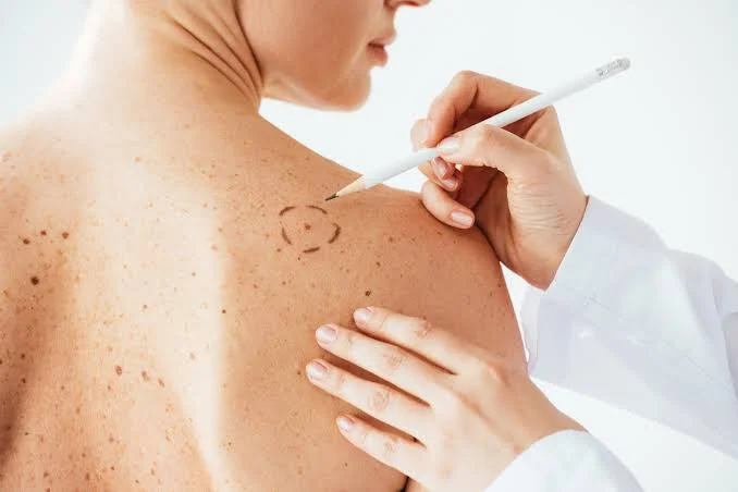
Signs of Melanoma: How to Spot Them Before It Spreads
Have you noticed a mole that looks slightly different and wondered whether you are overreacting? Most changing spots will not turn out to be cancer. Still, knowing the signs of melanoma can help you recognize a problem while it is smaller and easier to treat.
This disease does not always look like a large, dark mole. It may appear as a new freckle-like mark, a pink bump, a sore that keeps returning, or a line beneath a nail. Moreover, it can develop in places that rarely see sunlight.
This guide explains the ABCDE method, less familiar warning clues, high-risk body sites, self-check steps, and what happens during a professional examination. The aim is not to make every mole frightening. It is to make meaningful change easier to notice.
Why Early Recognition Matters
Current U.S. estimates project about 112,000 new cases in 2026. Recent SEER data report an overall five-year relative survival rate of 94.7%. However, stage makes a major difference: five-year relative survival is 100% for localized disease, 76% for regional disease, and 34% for distant disease.
These figures describe large groups, not one person’s future. Even so, they show why a suspicious change should be checked early instead of watched for many months.
early stage melanoma is often limited to its original site and may be treated with surgery alone. Once abnormal cells reach lymph nodes or distant organs, care usually becomes more complex.
One practical insight I would emphasize is simple: you do not need to know whether a spot is cancer before booking an appointment. Noticing that it is new, changing, or different is enough reason to ask a qualified clinician.
Signs of Melanoma and the ABCDE Rule
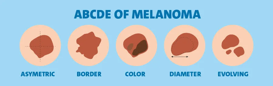
Dermatologists use the ABCDE rule to describe common warning patterns. It is useful for moles, freckles, and other pigmented spots, although it cannot diagnose cancer at home.
A Is for Asymmetry
Imagine drawing a line through the center. If one side does not match the other in shape, thickness, or color, the lesion is asymmetrical.
Many harmless moles are balanced. However, some healthy moles are not perfectly even, so consider this clue with the others.
B Is for Border
A concerning edge may be blurred, ragged, notched, scalloped, or hard to follow. Pigment may also appear to spread into nearby skin.
C Is for Color
Several colors inside one spot can require attention. These may include tan, brown, black, red, white, gray, pink, or blue.
A single color does not guarantee safety. amelanotic melanoma may contain little dark pigment and can look pink, red, pale, or skin-colored.
D Is for Diameter
Many suspicious lesions are larger than 6 millimeters, roughly the width of a pencil eraser. However, dangerous spots can be smaller.
Therefore, size should never be the only test. A tiny mark that is changing may deserve more attention than a larger mole that has remained stable for years.
E Is for Evolving
Evolution is often the most useful clue. Watch for changes in size, shape, height, color, border, texture, or feeling.
A lesion may also begin itching, crusting, bleeding, hurting, or forming a sore. The AAD advises medical review for a new spot, one that looks different, or one that changes, itches, or bleeds.
Signs of Melanoma Beyond an Unusual Mole
The ABCDE method does not describe every case. Some growths resemble cysts, scars, insect bites, warts, bruises, or ordinary sores.
nodular melanoma, for example, may appear as a firm, dome-shaped bump. It can be dark, red, pink, or skin-colored and may bleed after minor contact. Because it can grow downward quickly, waiting for every ABCDE feature may delay care.
Other warning changes include:
- A new spot that continues to enlarge
- A firm bump that feels different from nearby skin
- A sore that does not heal or heals and returns
- A mole that bleeds without a clear injury
- New itching, tenderness, crusting, or pain
- A scar-like patch without a past wound
- A dark streak or damaged area beneath a nail
The term malignant melanoma still appears in some records and older articles. It refers to the same serious cancer of pigment-producing cells, not a separate warning pattern.
Use the “Ugly Duckling” Clue
Most people’s moles share a general family resemblance. They may be similar in color, size, shape, or thickness.
An ugly duckling is one spot that looks noticeably different from the rest. For example, if nearly all your moles are small and light brown, a new black patch stands out. Likewise, when most are flat, a new firm bump deserves attention.
This approach is helpful for people who have many moles. Instead of expecting every lesion to match a textbook photograph, look for the one that breaks your normal pattern.
What Does It Look Like on Different Skin Tones?
Anyone can develop this disease. Fair skin, sunburn history, many unusual moles, family history, tanning-bed use, and heavy ultraviolet exposure can raise risk. However, people of every skin tone need to know their normal pattern.
In darker skin, suspicious growths may appear on the palms, soles, fingers, toes, or around a nail. They may also be found later when risk is underestimated.
The question what does melanoma look like on the skin has no single visual answer. A suspicious lesion can be black, brown, blue, red, pink, white, or almost colorless. Change and persistence matter more than one “classic” shade.
Remember to check the scalp, behind the ears, palms, soles, spaces between toes, buttocks, genital area, and beneath the nails.
Nail, Eye, and Other Less Obvious Locations
A dark nail streak can result from injury, natural pigment, medicine, or another harmless cause. Still, a new band on one nail that widens, darkens, damages the nail, or spreads onto nearby skin needs assessment.
People often search normal black line on nail vs melanoma after noticing a vertical stripe. Photographs can raise awareness, but only examination and, when required, biopsy can establish the cause.
subungual melanoma begins beneath or within the nail unit, while nail melanoma is a broader public term for tumors in this area. Warning changes can include splitting, lifting, a bump, bleeding, or dark skin around the nail.
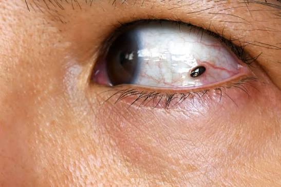
ocular melanoma begins in pigment-producing cells in or around the eye. New flashes, floaters, blurred vision, a missing area of sight, or a growing iris spot should be assessed by an eye specialist. It is biologically different from skin melanoma.
How to Perform a Skin Self-Exam
A regular check helps you learn what is normal for your body. People at higher risk may need a different schedule, so follow a dermatologist’s advice.
Choose a well-lit room, a full-length mirror, a hand mirror, and a phone camera. Remove nail polish and ask a partner to check difficult areas when possible.
Follow This Five-Step Check
- Face and scalp: Inspect the nose, lips, eyelids, ears, hairline, and scalp. Part the hair in small sections.
- Upper body: Check the neck, chest, stomach, shoulders, arms, elbows, underarms, hands, fingers, and nails.
- Back: Use a hand mirror for the back of the neck, shoulders, back, buttocks, and backs of the thighs.
- Lower body: Examine every side of the legs, then check the feet, heels, soles, toenails, and spaces between toes.
- Record changes: Photograph unusual spots in the same light and from the same angle. Add a date and, when possible, a ruler.
The AAD recommends examining the full body, including the scalp, back, soles, and between the toes—not only areas exposed to sunlight.
A body map can also help. Slow changes are easier to see when two dated images are compared side by side.
When Should You See a Dermatologist?
Arrange a professional examination when you find:
- A new or changing mole
- A spot unlike your others
- A lesion with ABCDE features
- A sore that does not heal
- Unexplained bleeding, crusting, pain, or itching
- A growing firm bump
- A new line or damaged area beneath a nail
- Any spot that concerns you despite not fitting a checklist
Do not repeatedly scratch, cut, burn, freeze, or treat a suspicious spot with home remedies. These actions can irritate the skin and delay diagnosis.
If cancer is suspected, a clinician may remove all or part of the lesion. The National Cancer Institute states that diagnosis requires tissue removal and examination for cancer cells.
What Happens During a Skin Examination?
The clinician will ask when the spot appeared, how it changed, whether it causes symptoms, and whether you have risk factors such as previous skin cancer, tanning exposure, severe sunburns, or family history.
Next, the skin is examined from the scalp to the feet. A dermatologist may use a dermatoscope, a lighted magnifying tool that shows structures not easily seen with the naked eye.
When a lesion is suspicious, a biopsy may be completed during the visit or scheduled soon afterward. A pathologist checks the sample and reports features such as tumor thickness and ulceration, which help guide staging and treatment.
Do not feel embarrassed to ask whether the examination will cover your whole body. A complete check may include the scalp, soles, nails, and other covered areas.
How Fast Can It Spread?
People frequently ask how fast does melanoma spread, but there is no universal timeline. Some lesions change slowly, while others grow deeper or reach nearby tissue more quickly.
Risk depends on tumor thickness, ulceration, growth pattern, location, and whether cells have reached lymph nodes or distant organs. That is why waiting many months can be unsafe when a spot is clearly evolving.
metastatic melanoma means the disease has reached distant parts of the body. New treatments have improved outcomes for many patients, but early diagnosis remains strongly linked with simpler care and better group survival.
Prevention and Common Detection Mistakes
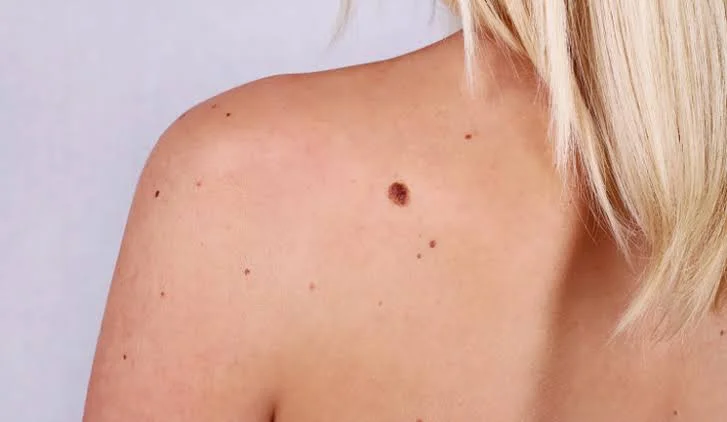
Reducing ultraviolet exposure can lower avoidable risk. Use broad-spectrum sunscreen, seek shade, wear protective clothes, and avoid tanning beds. However, prevention does not replace skin checks because suspicious lesions can appear in covered areas.
The phrase melanoma skin cancer is often used to separate cutaneous disease from eye or mucosal forms. Whatever the wording, remember four common mistakes: waiting for pain, expecting every lesion to be dark, ignoring spots smaller than 6 millimeters, and checking only sun-exposed skin.
A phone can document change, but it cannot replace an examination or biopsy.
What to Do After Finding a Suspicious Spot
Stay calm and take practical steps:
- Photograph the area clearly.
- Measure it with a ruler when possible.
- Write down when you first noticed it.
- Note itching, bleeding, pain, or recent growth.
- Avoid strong treatments before the appointment.
- Arrange medical review without waiting for another change.
For example, tell the clinic, “This spot appeared two months ago, has become darker, and now bleeds after light contact.” Specific information may help staff judge how quickly you should be seen.
Conclusion
Learning the signs of melanoma does not mean treating every mole as dangerous. It means knowing your usual pattern and responding when a spot is new, changing, different, bleeding, painful, or failing to heal.
Use the ABCDE method, but look beyond it. Check firm bumps, pink lesions, nail bands, unexplained scars, and hidden body areas. Moreover, document changes instead of relying only on memory.
Most examined spots are not cancer. Still, prompt evaluation offers reassurance when a lesion is harmless and faster treatment when it is not. In short, notice change, act early, and let a qualified dermatologist make the diagnosis.
Frequently Asked Questions
What is usually the first warning sign?
A change in an existing mole or a new, unusual spot is common. It may change in size, shape, color, surface, or feeling.
Can a suspicious spot be smaller than a pencil eraser?
Yes. Although many diagnosed lesions are larger than 6 millimeters, they can be smaller. Evolution and difference from your other spots are often more useful clues.
Does every unusual mole need a biopsy?
No. A dermatologist may decide that a spot is harmless or should be photographed and monitored. Biopsy is used when findings raise concern or the diagnosis remains uncertain.
Can a cancerous spot be pink?
Yes. Some lesions contain little visible pigment and can look pink, red, pale, or skin-colored. A growing or changing lesion should be checked regardless of color.
How often should I check my skin?
Many dermatology resources encourage regular self-exams, but the best schedule depends on personal risk. Previous skin cancer, atypical moles, or strong family history may justify more frequent professional checks.
⭐ Make MindLiva Your Preferred Source on Google
Get trusted health, wellness, and lifestyle articles from MindLiva.
Add MindLiva
Samie Babaei is a medical science student at Shahid Beheshti University of Medical Sciences with a strong interest in health, wellness, and medical research. As a health writer and researcher at MindLiva, she focuses on creating clear, evidence-based content that helps readers better understand health and well-being.