




Melanoma Cancer White Spots on Skin: Key Facts
Have you noticed a pale mark and immediately feared the worst? Searches for melanoma cancer white spots on skin often begin with that anxious moment.
Fortunately, most isolated white marks come from common pigment conditions, fungal overgrowth, dry skin, sun-related changes, or healing after irritation. They are not usually a sign of cancer.
However, color loss can sometimes appear within an unusual mole or patch. It may look uneven, scar-like, or mixed with brown, black, pink, red, gray, or blue.
In this guide, you will learn what white areas may mean, which changes deserve attention, how doctors examine suspicious spots, and why online photographs cannot provide a firm diagnosis.
Melanoma Cancer White Spots on Skin: The Clear Answer
A plain white spot by itself is not the usual appearance of this disease. A more common warning pattern is a new or changing lesion with an uneven shape, irregular edge, varied color, or steady growth.
The National Cancer Institute explains that suspicious lesions can contain areas of white or gray alongside brown, black, red, pink, or blue. Therefore, color should always be assessed as part of the whole lesion.
White or skin-colored areas may also appear when part of a pigmented lesion loses color. Dermatologists often describe this as regression or depigmentation.
The pale area may look uneven or scar-like rather than like a neat white freckle. However, this feature cannot confirm cancer because harmless skin conditions can also cause pigment loss.
In short, most white spots are not cancerous, but a pale area inside a changing or unusual lesion should be examined.
Why White Spots Usually Have Other Causes
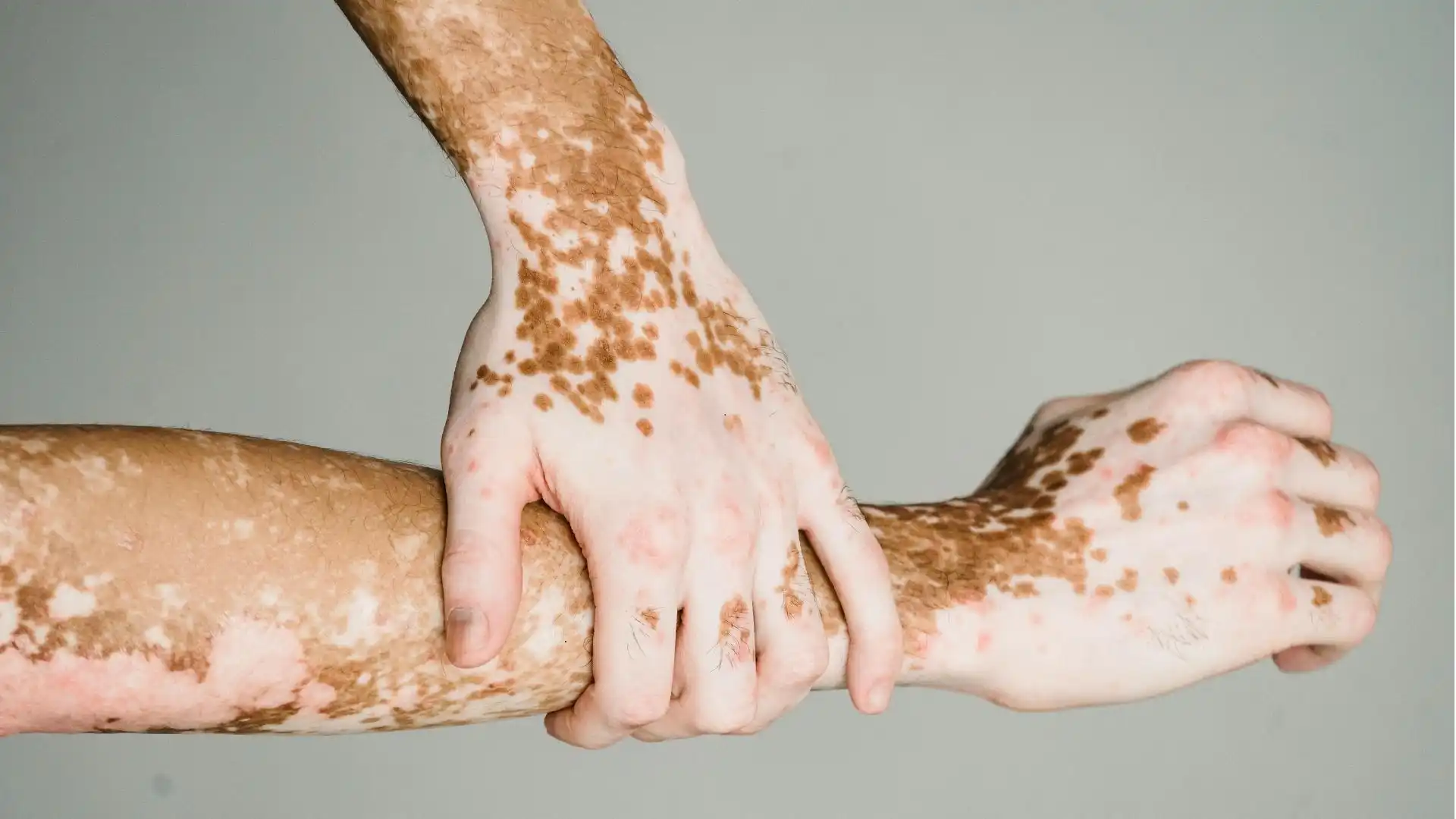
Skin pigment can decrease for many reasons. The location, shape, texture, speed of change, symptoms, and surrounding skin help a doctor identify the likely cause.
Vitiligo
Vitiligo causes one or more areas of skin to become lighter, white, or sometimes pink. It develops when pigment-making cells are destroyed.
The patches are commonly smooth and may expand over time. Hair growing from the affected area can also become white.
Unlike a suspicious mole, vitiligo usually creates a clearer area of color loss rather than a raised or irregular growth containing several colors. Nevertheless, an accurate diagnosis remains important.
Tinea Versicolor
Tinea versicolor is a common yeast-related condition. It may produce white, pink, tan, brown, or slightly darker patches.
These areas can be dry, mildly itchy, or scaly. They frequently develop on the chest, upper back, neck, abdomen, or upper arms.
The patches may become more noticeable after sun exposure because the affected area does not tan like the surrounding skin. Several small spots can also join and form a larger patch.
Post-Inflammatory Pigment Loss
A patch may become lighter after eczema, a rash, a burn, an insect bite, or another skin injury.
The original irritation may have disappeared by the time the pale mark becomes noticeable. This contrast is often more visible on brown or Black skin.
Small Sun-Related White Spots
Idiopathic guttate hypomelanosis causes small, flat, pale marks, commonly on sun-exposed forearms or lower legs. It is considered a benign form of pigment loss and becomes more common with age.
These spots are generally smooth, separate, and stable. However, a dermatologist can confirm the cause when the pattern is uncertain.
Pityriasis Alba and Dry Skin
Pityriasis alba can cause poorly defined pale patches with mild dryness or fine scaling. It commonly affects the face and upper arms and is often linked with dry or eczema-prone skin.
When a White Area Becomes More Concerning
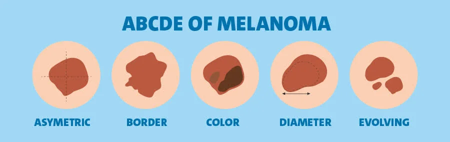
Do not judge a skin lesion by its white color alone. Instead, examine its entire pattern and consider whether it is new, different, or changing.
The ABCDE method provides a practical guide:
- A — Asymmetry: One half does not match the other.
- B — Border: The edge looks blurred, notched, ragged, or poorly defined.
- C — Color: Several shades appear within the same lesion.
- D — Diameter: The area may be larger than 6 millimeters, although smaller suspicious spots can occur.
- E — Evolving: The lesion changes in size, shape, color, height, texture, or sensation.
Another useful method is the ugly duckling rule. A mark that looks clearly different from every other spot on your body deserves attention, even when it does not meet every ABCDE feature.
Other signs of melanoma include bleeding, crusting, itching, tenderness, pigment spreading beyond the border, a sore that fails to heal, or a newly firm lump. These symptoms do not automatically mean cancer, but they justify professional assessment.
What Different Types Can Look Like
This disease does not have one fixed appearance. Its color and shape can depend on the subtype, location, depth, and amount of pigment present.
Flat or Slowly Expanding Lesions
A superficial lesion may begin as a flat patch that changes over months or years. It can contain brown, black, gray, blue, red, pink, skin-colored, or white scar-like areas.
When diagnosed as early stage melanoma, treatment and expected outcomes are generally more favorable than after deeper invasion or distant spread.
Current SEER data show that five-year relative survival is highest for localized disease. However, population statistics describe large groups and cannot predict one individual’s outcome.
Firm and Growing Bumps
Nodular melanoma often grows upward and may form a raised, firm, enlarging bump. It can appear dark, red, pink, or close to the surrounding skin color.
The **EFG guide—elevated, firm, and growing—**can help identify a lesion that does not fit the usual flat ABCDE pattern.
Lesions With Little Pigment
Amelanotic melanoma contains little visible dark pigment. It may resemble a pink or red sore, scar, cyst, irritated patch, or skin-colored bump.
Because color may not provide an obvious warning, continued growth, bleeding, firmness, and failure to heal become especially important clues.
The older phrase malignant melanoma is still used in some health content. Specialists now usually describe the exact subtype, thickness, stage, and other tumor features.
Hidden and Less Common Locations
Subungual melanoma begins beneath or around a fingernail or toenail. A dark band is more common than a white mark, but new or changing nail pigmentation should be assessed.
Warning features include a widening band, uneven color, nail damage, or pigment spreading onto nearby skin.
People searching normal black line on nail vs melanoma should remember that injuries, harmless pigment bands, medicines, and blood beneath the nail can also produce dark lines.
Nail melanoma cannot be safely confirmed or excluded from color or line width alone. A dermatologist may use medical history, dermoscopy, follow-up images, or a biopsy.
Ocular melanoma begins in the eye rather than on the outer skin. It requires assessment by eye-care and cancer specialists and should not be confused with ordinary white skin patches.
The phrase skin melanoma is sometimes used informally for cancer beginning in cutaneous pigment-producing cells.
Melanoma skin cancer can develop on both sun-exposed and protected areas, including the scalp, genitals, palms, soles, and beneath nails.
Metastatic melanoma describes disease that has spread to distant organs or tissues. That diagnosis depends on pathology and staging tests, not the appearance of a single surface mark.
Melanoma Cancer White Spots on Skin Across Skin Tones
White areas may be easier to notice on medium or deep skin tones, while redness may be harder to see. Therefore, checking only for a dark mole may cause someone to overlook other important changes.
Examine the palms, soles, scalp, behind the ears, between the toes, beneath the nails, and other areas that receive little direct sunlight.
People of every skin color can develop this cancer, and suspicious lesions can begin in either exposed or covered areas.
Personal insight: Compare a mark with your own normal skin pattern rather than relying only on a stock photograph. A lesion that is unique on your body may be more meaningful than one isolated feature.
How Doctors Evaluate White or Mixed-Color Spots
A dermatologist normally begins by asking several questions:
- When did the spot first appear?
- Has it changed in size, shape, or color?
- Does it itch, hurt, bleed, crust, or repeatedly reopen?
- Have you recently had a rash or injury?
- Is there a personal or family history of skin cancer?
The clinician then examines the spot and the surrounding skin. A dermatoscope may be used to reveal structures and pigment patterns that are difficult to see with the naked eye.
If the lesion remains suspicious, tissue is removed and examined by a pathologist. Biopsy is the only way to establish a definite diagnosis.
People commonly ask what does melanoma look like on the skin, but appearance varies too widely for one photograph to answer that question reliably.
Images can support awareness and help document change. However, an app, visual search, or online article cannot replace pathology.
How Quickly Should You Seek Medical Advice?

Arrange a routine medical appointment for a persistent white patch that spreads, scales, or has no clear cause.
Request a prompt dermatology assessment when a lesion:
- Changes over several weeks or months
- Looks unlike your other marks
- Contains several uneven colors
- Becomes raised or firm
- Bleeds, crusts, or fails to heal
- Develops an irregular border
- Causes new itching, pain, or tenderness
The question how fast does melanoma spread has no universal answer. Growth speed depends on the subtype, depth, ulceration, genetics, immune response, and other tumor features.
Some lesions progress slowly, while others grow more quickly. Waiting for pain is not a safe strategy because suspicious spots can be painless.
A Practical Five-Minute Skin Check
Use bright light, a full-length mirror, and a hand mirror. Ask someone you trust to inspect areas that are difficult to see.
- Check the face, lips, nose, ears, scalp, and neck.
- Examine the hands, arms, elbows, and underarms.
- Look at the chest, abdomen, sides, back, and buttocks.
- Inspect the legs, feet, heels, soles, and spaces between toes.
- Check every nail and compare new photographs with older images.
Avoid repeatedly scratching, bleaching, or treating an unexplained mark with strong home remedies. Irritation may change its appearance and make later assessment more difficult.
Why Early Review Matters in 2026
SEER estimates that the United States will record approximately 112,000 new cases and 8,510 deaths in 2026.
The same source reports a five-year relative survival rate of 100% for localized disease in its latest dataset. This figure is a population estimate rather than a guarantee for an individual patient.
Moreover, diagnostic apps and AI-based image tools are becoming easier to access. They may improve awareness, but they can also create false reassurance when users compare one mark with a single photograph.
The safest approach remains a professional examination followed by biopsy when medically necessary.
Conclusion
Most white marks have non-cancerous explanations, including vitiligo, tinea versicolor, dryness, inflammation, and age-related pigment changes.
However, a pale or scar-like area inside an uneven, changing, or multi-colored lesion deserves professional attention.
In short, change matters more than color alone. Monitor new marks, use the ABCDE and ugly-duckling methods, protect your skin from UV exposure, and seek medical advice when something looks different or continues to evolve.
Early action does not mean assuming the worst. It means replacing online uncertainty with a reliable medical answer.
Frequently Asked Questions
Are Small White Dots Usually Skin Cancer?
Usually not. Tiny, stable white marks are often related to harmless pigment changes. However, a growing, bleeding, uneven, or multi-colored spot should be examined.
Can a Cancerous Mole Contain White Areas?
Yes. Some suspicious lesions contain white, gray, red, pink, blue, brown, or black areas. A white scar-like section may reflect regression, but only an examination and biopsy can identify its cause.
Can Vitiligo Be Mistaken for Cancer?
Vitiligo typically produces smooth areas of pigment loss rather than an irregular growth. Still, medical assessment is appropriate when a patch is unusual, changing, or difficult to identify.
Should I Photograph a New White Spot?
Yes. A dated photograph beside a ruler may help document changes. However, photographs should support—not replace—a professional examination.
Does Sunscreen Remove Existing White Spots?
No. Sunscreen cannot restore lost pigment, but it can prevent tanning around pale areas and reduce further UV damage. Treatment depends on the underlying cause.
⭐ Make MindLiva Your Preferred Source on Google
Get trusted health, wellness, and lifestyle articles from MindLiva.
Add MindLiva
Samie Babaei is a medical science student at Shahid Beheshti University of Medical Sciences with a strong interest in health, wellness, and medical research. As a health writer and researcher at MindLiva, she focuses on creating clear, evidence-based content that helps readers better understand health and well-being.