



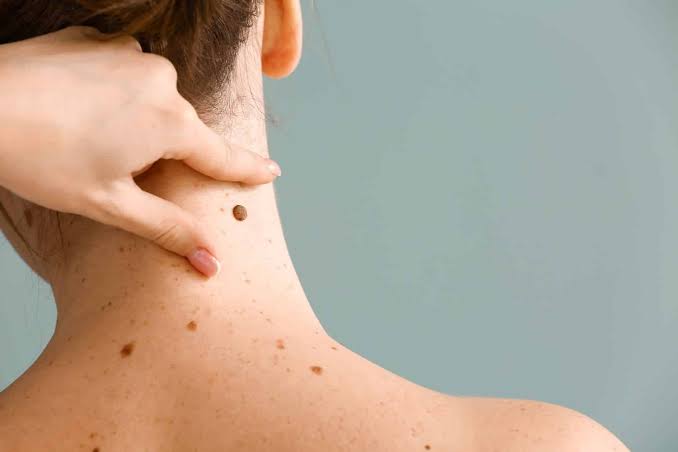
What Does Melanoma Look Like on the Skin? Key Visual Clues
Have you ever looked at a mole and wondered, “Could this be something serious?”
That question is worth asking because skin changes are common, while some can be early warning signs of cancer. Knowing what does melanoma look like on the skin can help you recognize changes that deserve professional attention.
Melanoma begins in melanocytes, the cells that produce melanin, the pigment responsible for much of our skin color. It can develop within an existing mole, but it can also appear as a completely new spot. The National Cancer Institute notes that changes in a mole’s shape, color, size, or feel are important warning signs.
In this guide, you will learn the most important visual clues, how the ABCDE rule works, why some melanomas are difficult to recognize, and when a dermatologist should examine a suspicious spot.
What Does Melanoma Look Like on the Skin? The Basic Clues
There is no single appearance that identifies melanoma.
Some lesions are dark and irregular. Others may be small, pink, red, or even similar to normal skin. Therefore, looking only for a classic black mole can lead to false reassurance.
The most important clues are usually change, asymmetry, irregular borders, uneven color, and an unusual appearance compared with other spots.
According to the American Cancer Society, a new spot or a spot that changes in size, shape, or color is one of the most important warning signs. A lesion that looks noticeably different from a person’s other moles—the so-called “ugly duckling” sign—can also deserve evaluation.
Common Visual Features
A suspicious lesion may:
- Have an uneven or asymmetrical shape
- Have an irregular or blurred border
- Contain several shades of color
- Become larger over time
- Change in texture or height
- Look noticeably different from surrounding moles
- Bleed, crust, or ooze
- Develop persistent itching or tenderness
However, none of these features alone proves cancer.
Many harmless moles can look unusual. The purpose of these signs is to help you decide when medical assessment is sensible.
The ABCDE Rule: A Simple Way to Check a Mole
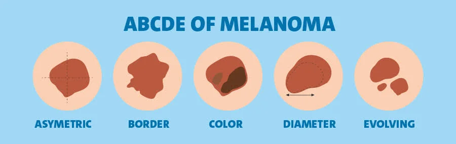
The ABCDE rule is one of the most useful tools for understanding what does melanoma look like on the skin.
It is an awareness guide, not a diagnosis.
A — Asymmetry
Imagine drawing a line through the center of a mole.
If one side looks very different from the other, the lesion is asymmetrical.
Normal moles are often relatively balanced in shape, although harmless moles can also have some irregularity.
B — Border
Look at the edge of the spot.
A concerning border may be:
- Ragged
- Notched
- Uneven
- Scalloped
- Poorly defined
- Blurred into the surrounding skin
The NCI specifically describes irregular, ragged, notched, or blurred borders as possible melanoma features.
C — Color
Healthy-looking moles often have a fairly even color.
A suspicious lesion may contain different shades, such as:
- Brown
- Black
- Tan
- Red
- Pink
- White
- Gray
- Blue
However, color alone is not enough.
Some benign lesions contain more than one shade, while some melanomas have surprisingly little pigment.
D — Diameter
The traditional ABCDE rule uses 6 millimeters—about the size of a pencil eraser—as a useful reference.
However, this number should never be treated as a strict cutoff.
A melanoma can be smaller than 6 millimeters, especially when it is detected early. The American Academy of Dermatology also emphasizes that melanomas can be smaller than this size.
E — Evolving
For many people, this may be the most important clue.
A mole that changes in:
- Size
- Shape
- Color
- Texture
- Height
- Symptoms
should be checked.
For example, a mole that looked the same for years but becomes darker and larger over several months deserves more attention than a stable mole that has looked unchanged for decades.
What Does Melanoma Look Like on the Skin Compared With a Normal Mole?
A normal mole is often:
- Symmetrical
- Evenly colored
- Smoothly bordered
- Relatively stable over time
Melanoma can be more irregular.
| Feature | Typical benign mole | Suspicious lesion |
| Shape | Often symmetrical | May be asymmetrical |
| Border | Usually smooth | May be irregular |
| Color | Often fairly uniform | May contain several colors |
| Size | Often stable | May enlarge |
| Change | Usually little change | May evolve noticeably |
| Appearance | Similar to other moles | May look like an “odd one out” |
This comparison is useful, but it is not a substitute for a clinical examination.
In real life, some lesions do not fit neatly into either column.
The “Ugly Duckling” Sign
Sometimes a lesion does not clearly satisfy every ABCDE rule.
Instead, it simply looks different from all the other moles around it.
This is called the ugly duckling sign.
For example, imagine someone has ten small, round, light-brown moles and one new spot that is much darker, larger, and irregular. Even if the unusual spot does not meet every ABCDE criterion, its difference may be important.
The American Cancer Society lists a spot that looks different from the others as a key warning sign.
This is especially useful for people with many moles because comparing lesions can sometimes be easier than judging each one in isolation.
Can Melanoma Look Like a New Mole?
Yes.
Melanoma does not always develop from a mole that has been present for years.
It may appear as a new pigmented or otherwise unusual area of skin.
This is one reason adults should pay attention to new spots.
A new mole is not automatically dangerous. Many new skin growths are harmless. However, a new lesion that continues to change should be evaluated rather than ignored.
What Color Is Melanoma?
Many people expect melanoma to be black.
It can be, but there is much more variation.
A suspicious lesion may contain:
- Dark brown
- Black
- Blue
- Gray
- Red
- Pink
- White
Sometimes several shades occur within the same lesion.
The NCI notes that melanoma can show combinations of black, brown, tan, white, gray, red, pink, or blue.
Therefore, uneven color is usually more meaningful than simply asking whether a mole is dark.
Can Melanoma Be Skin-Colored or Pink?
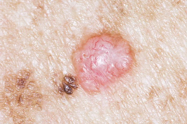
Yes.
Amelanotic melanoma produces little or no obvious dark pigment.
As a result, it may appear pink, red, flesh-colored, or pale.
This is one reason melanoma can be confused with other skin conditions.
For example, a persistent pink bump may look like irritation, an inflamed follicle, or another harmless growth. If it continues to grow, bleed, crust, or change, however, it should be examined.
This is also why photographs on the internet cannot provide a reliable diagnosis.
Nodular Melanoma Can Look Different
Nodular melanoma is a subtype that often appears as a raised growth rather than a flat, irregular patch.
It can be dark, but it may also appear pink, red, or flesh-colored.
Because this type can grow more deeply, a rapidly enlarging bump should not be ignored simply because it does not resemble a traditional dark mole.
A practical rule is simple: a new growth that changes quickly deserves medical attention.
Does Melanoma Itch or Hurt?
It can, but it does not have to.
A suspicious lesion may be completely painless.
In some cases, people report:
- Itching
- Tenderness
- Bleeding
- Crusting
- Oozing
- Sensitivity
The NCI notes that advanced lesions can become hard or lumpy and may ooze, bleed, itch, or become painful.
However, waiting for pain is not a good way to screen for cancer.
A painless but changing mole can still require evaluation.
Signs of Melanoma You Should Know
The signs of melanoma are broader than the classic ABCDE checklist.
Pay attention to a spot that:
- Appears suddenly
- Changes noticeably
- Grows over time
- Develops irregular color
- Has an unusual border
- Looks different from your other moles
- Bleeds or repeatedly crusts
- Changes in texture or height
A lesion does not need to have all eight features.
In fact, the NCI explains that some melanomas show only one or two ABCDE characteristics.
Early Stage Melanoma: Why Timing Matters
Finding early stage melanoma can make a major difference in treatment options.
When melanoma remains localized, surgery may be enough for some patients, depending on the tumor’s characteristics.
The American Academy of Dermatology emphasizes the value of finding melanoma early, when it is highly treatable.
This does not mean every suspicious spot is an emergency.
Instead, it means persistent or changing lesions should not be put off for months simply because they are not painful.
Early assessment gives a dermatologist the opportunity to determine whether a lesion is harmless or needs further testing.
What Does Melanoma Look Like on Different Parts of the Body?
Melanoma can occur almost anywhere on the skin.
On the:
- Face: It may be difficult to distinguish from sun spots or age-related pigmentation.
- Scalp: Hair can hide a changing mole.
- Back: A person may not notice changes without a mirror or assistance.
- Legs: New or changing moles can be easier to see.
- Feet: Lesions may be overlooked because they are hidden.
- Hands: Pigmented spots can sometimes be mistaken for minor injuries.
The American Academy of Dermatology recommends checking areas that are easy to overlook, including the scalp, back, nails, soles, and spaces between the toes.
Melanoma on the Scalp: A Hidden Risk
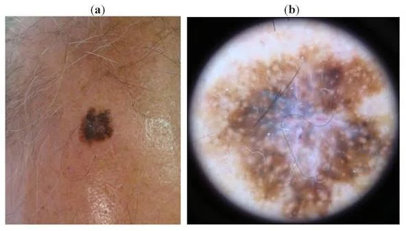
Scalp lesions deserve special attention because hair can conceal them.
A person with thick hair may never see the skin underneath unless they deliberately part the hair.
Try using a handheld mirror or asking someone you trust to check difficult areas.
Look for a new mole, an enlarging spot, uneven pigmentation, bleeding, or a lesion that looks different from nearby skin.
What About Nail Melanoma?
Melanoma can also develop under a nail.
Subungual melanoma occurs beneath the nail and may initially appear as a brown or black vertical streak.
People often search normal black line on nail vs melanoma because ordinary nail pigmentation and cancer can look similar.
A dark streak does not automatically mean cancer. Trauma, medications, benign pigmentation, and other conditions can cause nail discoloration.
However, a new or changing streak should be assessed by a healthcare professional.
Nail melanoma is uncommon, but delayed recognition can occur because the change is sometimes mistaken for an injury.
Understanding Malignant Melanoma
Malignant melanoma refers to melanoma that has the potential to invade tissue and spread.
That sounds alarming, but prognosis varies widely.
Doctors consider factors such as:
- Tumor thickness
- Ulceration
- Lymph node involvement
- Whether distant organs are affected
- Tumor biology
- Overall stage
Therefore, a diagnosis alone does not tell you exactly how serious a particular case will be.
The earlier a suspicious lesion is assessed, the sooner appropriate treatment can be considered.
How Fast Does Melanoma Spread?
Patients frequently ask, how fast does melanoma spread?
There is no single answer.
Different melanomas behave differently. Some can progress quickly, while others develop more slowly.
Tumor thickness, subtype, location, genetic features, immune response, and stage can all influence behavior.
For this reason, doctors do not recommend using time alone to judge whether a spot is safe.
A changing lesion should be evaluated rather than monitored indefinitely at home.
What Happens When Melanoma Spreads?
If cancer moves from the original skin site to distant parts of the body, it may be called metastatic melanoma.
Treatment at this stage can include immunotherapy, targeted medicines, surgery, radiation, or combinations of treatments depending on the patient’s situation.
Modern treatment has greatly expanded options for advanced disease.
Still, the best strategy remains individualized care based on staging and tumor characteristics.
Other Types You May Hear About
Understanding the terminology can prevent confusion.
Ocular melanoma begins in the eye and is different from most skin tumors.
Subungual melanoma develops beneath a fingernail or toenail.
Amelanotic melanoma has little visible pigment and can be pink, red, or skin-colored.
These are all related to melanocytes, but their appearance, diagnosis, biology, and treatment can differ.
Likewise, skin melanoma is a broad term for melanoma arising in the skin.
How Doctors Confirm a Suspicious Mole
A dermatologist may begin with a visual examination and dermoscopy.
Dermoscopy provides magnified views of structures beneath the skin surface and can help the doctor decide whether a lesion looks benign or suspicious.
However, the only way to definitively diagnose melanoma is to examine tissue under a microscope after a biopsy. The NCI explicitly states that tissue examination is required for diagnosis.
Why You Should Not Remove a Mole at Home
Trying to cut, burn, freeze, or otherwise remove a mole yourself can cause:
- Infection
- Scarring
- Bleeding
- Incomplete removal
- Delayed diagnosis
A suspicious mole should be assessed professionally.
How to Check Your Skin at Home
A regular self-exam can help you notice changes.
The American Academy of Dermatology recommends using mirrors to inspect difficult areas and specifically advises checking the scalp by parting the hair.
A Practical Skin-Check Routine
Step 1: Stand somewhere with good lighting.
Step 2: Examine your face, neck, arms, hands, and torso.
Step 3: Check your legs, feet, soles, and between your toes.
Step 4: Inspect your scalp by separating the hair.
Step 5: Examine your nails.
Step 6: Use a mirror or ask someone to help with your back.
Step 7: Compare suspicious spots with older photographs when available.
Step 8: Contact a dermatologist about anything new or changing.
A partner can also help check areas that are difficult to see. The AAD notes that partners can help identify new melanomas during skin examinations.
Personal Insight: Look for Change, Not Perfection
One of the most useful lessons when checking your skin is that you do not need to become an expert at identifying cancer.
Instead, learn what is normal for you.
Imagine that you have several small, evenly colored brown moles. Then, one month later, you notice a new dark spot that is much larger and has an uneven border.
You do not need to decide whether it is cancer.
You only need to recognize that it is different and arrange an appropriate medical examination.
That approach is practical, less stressful, and much safer than trying to diagnose yourself from photographs.
Can You Prevent Melanoma?
No prevention method eliminates the risk completely.
However, reducing unnecessary UV exposure is an important part of skin cancer prevention.
Helpful habits include:
- Wear protective clothing in strong sunlight.
- Use broad-spectrum sunscreen on exposed skin.
- Reapply sunscreen as directed.
- Seek shade when UV levels are high.
- Avoid indoor tanning.
- Wear a hat that protects the face, ears, and scalp.
- Check your skin regularly.
Sun protection is especially important for people with a history of intense sun exposure or sunburn.
Melanoma Skin Cancer vs. Other Skin Problems
Many conditions can resemble melanoma.
For example, a dark spot could be a benign mole, seborrheic keratosis, sun-related pigmentation, or another harmless growth.
Likewise, a red or pink bump could have many causes unrelated to cancer.
This is why online image searches have limitations.
Even experienced clinicians sometimes need dermoscopy or a biopsy to determine what a lesion actually is.
If you are unsure about a changing lesion, professional assessment is more useful than repeatedly comparing it with internet pictures.
When Should You See a Dermatologist?
Make an appointment if you notice a spot that is:
- New and unusual
- Changing in size
- Changing in shape
- Changing in color
- Bleeding
- Crusting repeatedly
- Itching persistently
- Growing quickly
- Different from your other moles
You should also seek advice if a spot simply looks unusual enough to concern you.
There is no benefit in trying to “wait and see” for months when a professional can examine it.
The Role of Technology in Melanoma Detection
Digital photography and dermoscopy have made it easier to track suspicious lesions.
Some clinics use total-body photography or sequential digital dermoscopy to monitor patients with many moles.
Artificial intelligence is also being studied as a support tool for skin cancer detection.
However, the American Cancer Society notes that consumer AI tools still have accuracy concerns and should not replace examination by a trained healthcare professional.
Technology can support medical care, but it should not become a substitute for it.
What Does Melanoma Look Like on the Skin? The Key Takeaway
So, what does melanoma look like on the skin?
It may look like a new or changing mole, an asymmetrical spot, an irregularly bordered lesion, or an area with uneven colors. It may also be small, pink, red, or skin-colored.
The most important clue is often evolution.
A lesion that changes should get attention, even when it does not look like a textbook example.
Remember, though, visual clues cannot diagnose cancer. Only a healthcare professional—and, when needed, a biopsy—can provide a definitive diagnosis.
Conclusion: Know Your Skin, Notice the Changes
Understanding what does melanoma look like on the skin is less about memorizing one frightening image and more about learning to recognize meaningful changes.
Look for asymmetry, irregular borders, uneven colors, growth, and especially evolution. Also remember that some melanomas do not look dark or dramatic.
The best habit is simple: know what your skin normally looks like and investigate changes that persist or evolve.
Most unusual spots are not cancerous. However, checking them is far better than ignoring a change that later proves important.
In short, you do not need to diagnose a suspicious spot yourself. You simply need to notice when something has changed and know when to ask a professional.
FAQs: What Does Melanoma Look Like on the Skin?
What is the most common visual sign of melanoma?
A new or changing spot is one of the most important warning signs. Changes in size, shape, color, or texture should be evaluated by a healthcare professional.
Can melanoma be smaller than 6 millimeters?
Yes. Although the ABCDE rule uses 6 millimeters as a useful reference, melanoma can be smaller. A changing small lesion should not be dismissed because of its size.
Can melanoma be pink instead of black?
Yes. Amelanotic or lightly pigmented melanomas may appear pink, red, or skin-colored. This is one reason color alone cannot rule out melanoma.
Does a painful mole always mean melanoma?
No. Pain can have many causes, and melanoma can also be painless. Changes in appearance, growth, bleeding, or persistent symptoms are more useful reasons to seek an examination.
Can I diagnose melanoma from a photo?
No. Photos can help you notice changes, but they cannot provide a reliable diagnosis. A dermatologist may use dermoscopy and, when necessary, a biopsy to confirm what a lesion is.
⭐ Make MindLiva Your Preferred Source on Google
Get trusted health, wellness, and lifestyle articles from MindLiva.
Add MindLiva
Samie Babaei is a medical science student at Shahid Beheshti University of Medical Sciences with a strong interest in health, wellness, and medical research. As a health writer and researcher at MindLiva, she focuses on creating clear, evidence-based content that helps readers better understand health and well-being.